2984
SNR Comparisons of the Coils used for Neonatal Imaging1Diagnostic Imaging and Radiology, Childrens National Medical Center, Washington, DC, United States
Synopsis
Neonatal brain MRI is widely used in clinical and research scans, in which images are acquired with adult head coils or special neonatal head coils. However, the performances of these coils are not very clear. In this work, we provided detailed comparisons of the signal-to-noise ratio with five noise measurement methods, among adult 8-channel head coil, adult 32-channel head coil, and neonatal 8-channel head coil. The results suggested that adult 8-channel coil has the highest signal-to-noise ratio.
INTRODUCTION
Neonatal brain MRI is used clinically to detect the brain lesions and to assess central nervous system development. Although conventional brain receiver coils are designed for adult brain imaging, adult brain coils are widely used to in the neonatal brain MRI, due to the limited availability of the neonatal coils. There also exist commercial coils designed specifically for the neonatal brain. These have a small inner volume for improved SNR and coil sensitivity but tend to have less channels compared to some adult coils (e.g. 32 channels). However, coil performance is not only determined by the coil size or the number of channels, and the performance of these different coils in neonatal MRI has not been compared in details. Therefore, the goal of this study was to identify which coil would provide the highest signal-to-noise (SNR) in neonatal MRI at 3T.
Methods
To remove motion artifact as a factor influencing SNR, we used an in-house built phantom that mimicked the composition of the GE ‘braino’ spectroscopy phantom and was cylindrical in shape for this study.
The scans were performed on a GE Discovery 750 3T scanner. The coils that we used in this study included the adult 32-channel head coil (denoted by Adult32), the adult 8-channel head coil (denoted by Adult8), and the neonatal 8-channel head coil (denoted by Neonatal) from the LMT Medical Systems GmbH, Germany. Single shot fast spin echo was acquired from one single slice with thickness 10mm, which provided sufficient SNR. FOV was 160mm and the acquired matrix was 160x160. To simplify the problem, no parallel imaging was used. Readout bandwidth was ±25KHz. The minimum TE and the minimum TR were used.
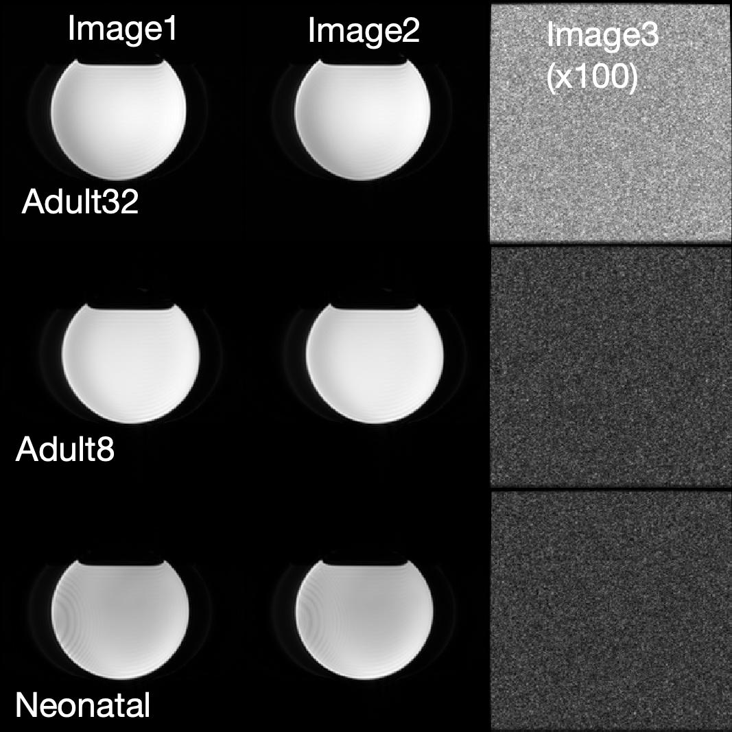
For each coil, we acquired three images (denoted by Image1, Image2, and Image3). Image1 and Image2 were regular images acquired repeatedly. Image 3 was acquired using the same imaging protocol with all RF pulses turned off. All images were acquired using the same prescan parameters to keep the same receive/transmit gains, and with one-minute delay after the prescan to achieve fully recovered magnetization.
Dicom images reconstructed online were processed in MATLAB 2018a. Because zero pixels was introduced during the distortion correction, the outer layer of the images background was removed.
Given that an accurate SNR measurement is based on the measurement of noise, we used five methods to estimate noise:
1. Noise1: the standard deviation of background in the Image2, which is commonly used for noise measurement
2. Noise2: based on the difference image between Image1 and Image2, the standard deviation of pixels was measured in the object region with a compensation factor of $$$\sqrt{2}$$$
3. Noise3: similar to Noise2, but the noise was measured from the background region1
4. Noise4: the standard deviation of pixels in Image3
5. Noise5: the mean square of pixels in Image3 with a compensation of the number of coils2
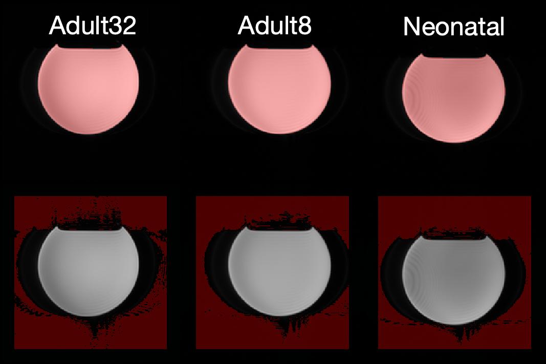
The object regions were selected as the pixels were larger than 500*Noise5 and the background regions were selected as the pixels were smaller than 10*Noise5.
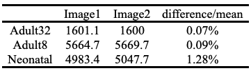
The signal was measured as the mean of the pixel values in the object region. The SNR was calculated as the mean of the signal between Image1 and Image2 scan divided by the noise defined above.
Results
The acquired images, scaled by the Image2, and selections of the regions are shown in figures 1 and 2. Table 1 shows the signal in the object regions, which had negligible differences between scans.
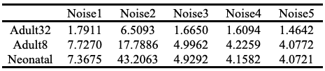
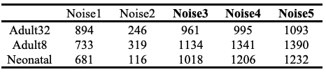
The measured noise and SNR were listed in table 2 and 3. The SNR values have dramatic differences depending on the measurements/definitions of noises. Comparing the SNR among coils, the Noise3, Noise4, and Noise5 showed the consistent results that adult 8-channel head coil provided highest SNR and adult 32-channel head coil resulted in the lowest SNR.
Discussion
The absolute SNR measurement highly depends on the definition and measurements of the noise. In this work, three out of five measurements showed that the adult 8-channel head coil provided the best SNR, which suggests the use of this coil for neonatal MRI.Acknowledgements
This work was partly supported by R01HL116585 from NIH National Heart, Lung, and Blood InstituteReferences
1. Dietrich O, Raya JG, Reeder SB, Reiser MF, Schoenberg SO. Measurement of signal-to-noise ratios in MR images: Influence of multichannel coils, parallel imaging, and reconstruction filters. J. Magn. Reson. Imaging 2007;26:375–385 doi: 10.1002/jmri.20969.
2. Constantinides CD, Atalar E, McVeigh ER. Signal-to-noise measurements in magnitude images from NMR phased arrays. Magn. Reson. Med. 1997;38:852–857 doi: 10.1002/mrm.1910380524.
Figures