2981
Accelerated Compressed Sensing 3D multi-parametric imaging Toward Isotropic1mm3 Imaging1MR Applications and Workflow, GE Healthcare, Tokyo, Japan, 2Radiology, Juntendo University School of Medicine, Tokyo, Japan, 3Radiology, The University of Tokyo Graduate School of Medicine, Tokyo, Japan, 4Department of Imaging Physics, The University of Texas M.D. Anderson Cancer Center, Houston, TX, United States, 5SyntheticMR, Linkoping, Sweden, 6MR Applications and Workflow, GE Healthcare, Menlo Park, CA, United States, 7ASL Europe, GE Healthcare, Munich, Germany, 8Juntendo University School of Medicine, Tokyo, Japan
Synopsis
A multi-parametric technique, 3D QALAS, accelerated using compressed sensing, was implemented to yield images of the brain at high spatial resolution of 1mm isotropic with multiple contrast weightings as well as parametric maps from a single scan. Compressed sensing factor of 1.5 combined with parallel imaging was used to accelerate scan time. The proposed 3D technique is expected to achieve both stable image quality and tissue segmentation accuracy with short imaging time.
Purpose
MAGiC1 is a promising 2D multi-contrast MRI technique that can obtain both multiple contrast images and high spatial resolution in a short imaging time. However, this has some limitations. For example, it might be necessary to repeat the acquisition in another plane for better visualization of pathology and high spatial resolution isotropic voxel acquisition is desired for brain morphometry measurement2. 3D QALAS3-4, or so called a version of 3D MAGiC was recently developed to perform volumetric high-resolution imaging of the brain and to reduce the partial volume effect of the tissue boundary, improving accuracy of parametric mapping. In this work, we applied compressed sensing (CS) to the QALAS and performed a feasibility study to achieve isotropic voxels of 1 mm with higher accelerated imaging.Methods
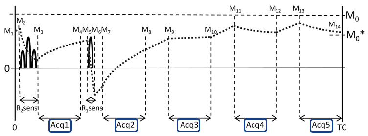
3D QALAS obtains five image contrasts using 3D gradient echo sequence in Fig.1. The first acquires MR data after T2 preparation pulse. The second and later are performed during the following T1 relaxation after Inversion pulse. Five data collections are executed at equal time intervals of 1 second. The 3D data is acquired with segmented Cartesian k-space sampling. 100 phase encodings are acquired per segment, and the sequence is repeated until enough segments are acquired to fill k-space. Quantitative T1, T2 and PD maps were reconstructed from the obtained images using a research version of SyMRI (SyntheticMR, Linkoping, Sweden). White matter(WM), gray matter(GM) and CSF volume segmentation of intracranial volume (ICV) were also performed5.
For compressed sensing, serial combination of CS + parallel imaging (ARC) was used6. ARC parallel imaging7 acceleration was fixed at factor 2. CS acceleration factor was varied between 1.3, 1.5 and 1.8. In order to evaluate the differences between the receiver coils, data were acquired with both 12 channel and 32 channel receiver coils. Uniformly undersamped k-space points by parallel imaging were retrospectively randomly undersampled to simulate CS undersampling. Structural Similarity Index Measurements(SSIM) 8 and Peak Signal-to-Noise Ratio (PSNR) were calculated for reconstruction quality. For the reference image, image with ARC reduction factor of 2 was used. Comparisons were made for each axial section/slice against the reference image, and measurements were computed over the whole brain. Healthy volunteer scan was performed at 1 mm iso resolution while applying a CS factor of 1.5, using 12 channel coils (GEM HNU, GE Healthcare) and 32 channel coils (MR instrument) on a 3.0T System (Discovery MR 750w, GE Healthcare). Imaging parameters were: spatial resolution 1mm3, FOV = 25.6x20.5x14.6 cm, Matrix = 256x205x146, TR/TE 8.6/3.5 ms, bandwidth 25kHz, FA 5 degree. Scan time was 11:41 with ARC acceleration factor=2, and 7:41 for when an additional CS factor =1.5 was applied.
Results
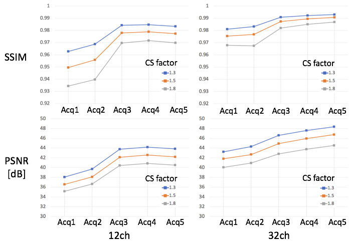
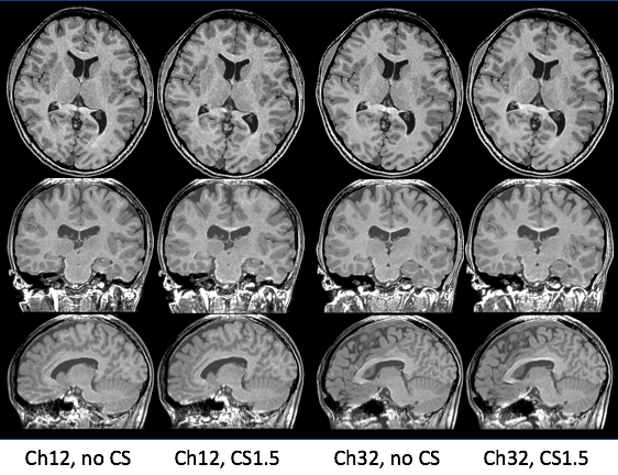
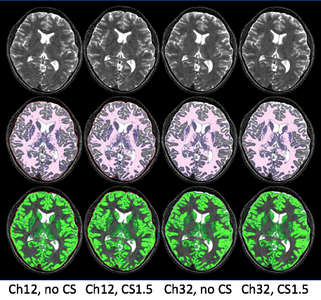
Fig.2 shows the results of SSIM and PSNR measurement by simulation. When CS factor was increased, both SSIM and PSNR showed progressively lower values suggesting deteriorating image quality. Image quality was lower in the early acquisitions of 3D QALAS compared to the later acquisitions, which maintained high values of SSIM and PSNR in both 12 and 32 channel. The 32 channel image maintained higher image quality than the 12 channel image when CS acceleration factor was increased. Fig.3 shows the result of T1 weighted volumetric image reconstructed by the syMRI. The image of CS acceleration factor 1.5 was visually similar compared to that of no CS acceleration in both 12 and 32 channel image. Fig.4 shows T2 weighted image and volume segmentation of the WM and the GM. The segmentation of the WM and the GM in CS factor of 1.5 showed similar result to no CS acceleration in both 12 and 32 channel. The WM volume fraction in relation to the ICV were 41.0% for no CS of the 12 channel, 41.1% for CS factor of 1.5 of the 12 channel, 42.3% for no CS of the 32 channel, 42.8% for CS factor of 1.5 of the 32 channel, corresponding to the GM fraction of 45.4%, 45.4%, 44.1%, 43.2% and the CBF fraction of 9.1%, 8.7%, 9.2% and 8.8%, respectively.
Discussions and Conclusion
We successfully demonstrated a 3D QALAS based multi-parametric technique in the brain that was accelerated using CS while maintaining image quality and tissue segmentation accuracy. As seen by the comparison between 12 and 32 channel coils, the increased SNR of higher multi-channel coils can be utilized to achieve both higher speed and stable image quality. Indicated by the simulation result, careful selection of CS factor would be necessary as the original image of each acquisition of QALAS deteriorates differently by CS factor. Although parallel imaging acceleration was fixed at factor of two in this study, optimized combination of ARC and CS factor could produce further acceleration of scan time.Acknowledgements
No acknowledgement found.References
1. Warntjes JB, Leinhard OD, West J, Lundberg P. Rapid magnetic resonance quantification on the brain: Optimization for clinical usage. Magn Reson Med. 2008 ; 60:320-9.
2. Jack CR Jr, Bernstein MA, et al. The Alzheimer's Disease Neuroimaging Initiative (ADNI): MRI methods. J Magn Reson Imaging. 2008 Apr;27(4):685-91.
3. Simultaneous three-dimensional myocardial T1 and T2 mapping in one breath hold with 3D-QALAS.Kvernby S, Warntjes MJ, Haraldsson H, Carlhäll CJ, Engvall J, Ebbers T.J Cardiovasc Magn Reson. 2014 Dec 20;16:102. doi: 10.1186/s12968-014-0102-0.
4. Hwang et al, Proc. Intl. Soc. Mag. Reson. Med. 18 (2019), 5627.
5. Warntjes M, Engström M, Tisell A, Lundberg P. Modeling the Presence of Myelin and Edema in the Brain Based on Multi-Parametric Quantitative MRI. Front Neurol. 2016 Feb 17;7:16.
6. King et al, Proc. Intl. Soc. Mag. Reson. Med. 18 (2010), 4881.
7. Anja, Brau et al, MRM 2008;59:382.
8. Zhou W, Bovik AC, Sheikh HR, Simoncelli EP. Image quality assessment: from error visibility to structural similarity. IEEE Transactions on Image Processing 2004;13(4):600-612.
Figures