2966
The value of DWI in the pre-treatment prediction of progression in Nasopharyngeal Carcinoma based on MRI radiomics1Department of Diagnostic Radiology, Cancer Hospital, Chinese Academy of Medical Sciences, Shenzhen Center, Shenzhen, China, 2GE Healthcare, MR Research China, Beijing, China, 3Huiying Medical Technology Co., Ltd., Beijing, China
Synopsis
Radiomics provides a new evaluation method for the prognosis of nasopharyngeal carcinoma (NPC) by extracting high throughput of quantitative descriptors from routinely acquired medical images. However, the research of the prognosis prediction of NPC based on radiomics derived from DWI images hasn’t been reported yet. The present study explores the value of DWI images in the pretreatment predictive of NPC with radiomics methods. By comparing the performance of three classifiers and different MRI sequences, we found that combined DWI and T2W showed optimal pretreatment predictive performance of NPC.
Introduction
Nasopharyngeal carcinoma (NPC) is the common malignant tumor in southeast China. In the recent years, the survival rates of NPC have been highly improved because of the radiotherapy technology. However,the prognosis assessment is still very important to avoid treatment failure of local recurrences and distant metastasis, especially for the patients with high grade NPC.1 Radiomics enables the noninvasive profiling of tumor heterogeneity, which make it more suitable in prognosis prediction of NPC.2 It has been reported that radiomics have shown great potential in tumor diagnosis, treatment and prognosis.3-5 In previous researches, radiomic features derived from joint axial T2-weighted image (T2W) and contrast-enhanced T1-weighted axial image (T1W_AX_C) were significant associated with progression free survival (PFS) of stage III-IVb NPC patients.1 However,the studies have omitted the value of diffusion weighted imaging (DWI) to the radiomic features of the pre-treatment prediction of progression of NPC. And the radiomic features of DWI have been reported to be significant in the tumor diagnosis.6,7 Therefore, this study aims to explore the value of DWI in the progression prediction of NPC.Methods
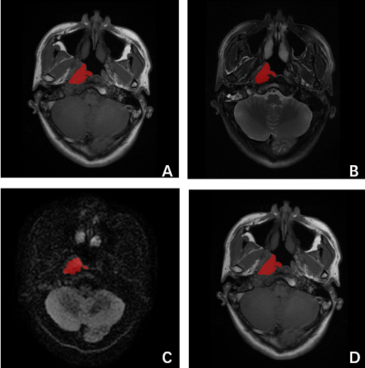
A total of 101 patients with diagnosed Stage I-IVB NPC were enrolled in this retrospective study. All the patients with no previous NPC treatment history, and whom were underwent nasopharynx and neck MRI examination (Discovery MR750, GE, WI) from Dec 13th, 2012 to Dec 23th, 2015 in Cancer Hospital, Chinese Academy of Medical Sciences. We chose the progression as the clinical endpoint. Patients were labeled as 1 if its progression-free survival (PFS) value smaller than 3 years, whereas the patients were labeled as 0 if its PFS value bigger than 3 years. The digital imaging and communications in medicine (DICOM) images including axial T1-weighted (T1W), T2WI, DWI and T1WI_AX_C, which were obtained from the PACS without normalization, which were used for segmentation, feature selection and the prognosis prediction of NPC. The open source software ITK-SNAP is used for 3D tumor segmentation manually, and the region of interest (ROI) was delineated on each slice of axial T1W, T2W, DWI and T1W_AX_C, which is shown in Fig.1. After the tumor segmentation, the radiomics feature extractor was adopted to extract all the features including the shape, signal intensity and texture (namely gray-level co-occurrence matrices (GLCM), gray level run length matrix (GLRLM) and gray level size zone matrix(GLSZM)) of the original imaging data of the lesions, as well as the signal intensity and texture features after processing through wavelet transformation, square root filtering, square filtering, exponential filtering and logarithmic filtering. Then, least absolute shrinkage and selection operator (Lasso) method was used to select the features most significant to the prognosis prediction of NPC on each images sequence and the joint images sequences respectively. The radiomics-based models were constructed with the selected features. And three classifiers, logistic regression (LR), random forest (RF), and support vector machine (SVM) with 5-fold cross-validation method were used to analyze the predict progression of NPC respectively. Finally, the receiver operating characteristic curve, namely, area under curve (AUC) and accuracy (ACC) were used to assess the predictive performance.Results and Disscusion
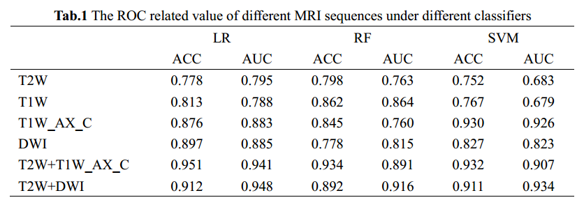
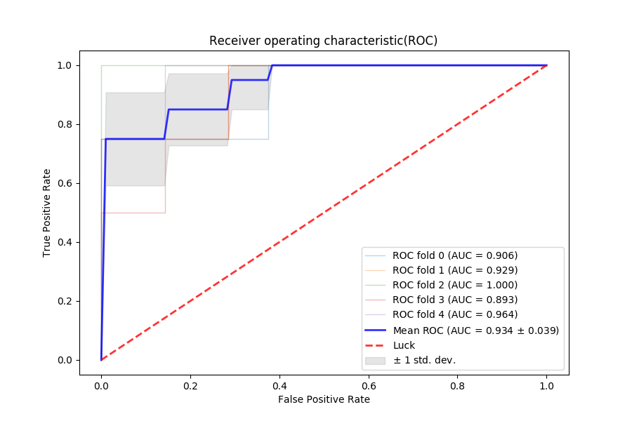
Results of the ROC related ACC and AUC value of different MRI sequences under LR, RF and SVM classifier respectively were summarized in Tab.1. Comparing with T2W, T1W and DWI, the T1W_AX_C (ACC=0.930, AUC=0.926) was shown to have the best predictive value under SVM. Meanwhile, the result of DWI (ACC=0.827, AUC=0.823) was more closer to T1W_AX_C than T2W and T1W, which indicate the potential predictive value of DWI. Then we combined T2W with T1W_AX_C and DWI respectively, the results indicated that T2W+DWI (AUC=0.934) had greater predictive value than T2W+T1W_AX_C. Fig.2 showed the ROC curve of T2W+DWI with SVM. Our result proved that the radiomics-based model on the joint T2W+T1W_AX_C sequences had high accuracy in the pre-treatment progression prediction which was reported in previous research.1 Our result also demonstrated that DWI has great potential in progression prediction in NPC patients before treatment, especially under the combination with T2W, and which can have the same predictive value as T1W_AX_C or even better. Since the DWI related radiomics research still remain relatively scarce, more evidence need to be put forward. In the following stage, we will enroll more cases into research and find more support.Conclusion
DWI and T2W+DWI based radiomic features have great value or high accuracy in the pre-treatment prediction of progression in NPC, especially T2W+DWI.Acknowledgements
No acknowledgement found.References
1. Zhang B, Tian J, Dong D, et al. Radiomics Features of Multiparametric MRI as Novel Prognostic Factors in Advanced Nasopharyngeal Carcinoma [J]. Clinical cancer research : an official journal of the American Association for Cancer Research, 2017, 23(15): 4259-69.
2. Gillies RJ, Kinahan PE, Hricak H. Radiomics: Images Are More than Pictures, They Are Data [J]. Radiology, 2016, 278(2): 563-77.
3. Scrivener M, de Jong EEC, van Timmeren JE, et al. Radiomics applied to lung cancer: a review [J]. Translational Cancer Research, 2016, 5(4): 398-409.
4. Chaddad A, Sabri S, Niazi T, et al. Prediction of survival with multi-scale radiomic analysis in glioblastoma patients [J]. Medical & Biological Engineering & Computing, 2018.
5. Kickingereder P, Neuberger U, Bonekamp D, et al. Radiomic subtyping improves disease stratification beyond key molecular, clinical, and standard imaging characteristics in patients with glioblastoma [J]. Neuro-Oncology, 2018, 20(6): 848-57.
6. Brown AM, Nagala S, McLean MA, et al. Multi-institutional validation of a novel textural analysis tool for preoperative stratification of suspected thyroid tumors on diffusion-weighted MRI [J]. Magnetic resonance in medicine, 2016, 75(4): 1708-16.
7. Fruehwald-Pallamar J, Czerny C, Holzer-Fruehwald L, et al. Texture-based and diffusion-weighted discrimination of parotid gland lesions on MR images at 3.0 Tesla [J]. NMR in biomedicine, 2013, 26(11): 1372-9.
Figures