2964
An investigation of the spatial FOV fold-over oversampling effects in the phase signal of flow-encoding cerebral 2D Cine PC-MRI: an application in phantom and volunteers.1MRI Research GIE-FF, CHU Amiens Picardie, Amiens, France, 2Pole Imagerie, CHU Amiens Picardie, Amiens, France
Synopsis
Acquiring high spatial resolution cine phase contrast MRI to quantify the blood flow (arteries or veins) and the cerebrospinal fluid flow (aqueduct or cervical) is challenging. The need for large number of voxels in such small section area is required to accurately quantify the flows measurements. Reduced FOV with fold-over suppression instead of spatial pre-saturation slab is an alternative to such application. The purpose of this study is to investigate the effect of oversampling option in the VNR and both the blood and CSF flow velocities measurements ex-vivo and in-vivo.
Introduction
Cine PC-MRI provides quantitative cerebro-spinal fluid (CSF) and blood flow velocity, peak velocity and other dynamic parameters that can be used to grade the severity of hydrocephaly and cerebro-vascular dysfunctions. CSF and blood flow visualizations have been successfully performed in various parts of the human brain including the aqueduct; the foramen of Magendie [1], arteries and veins [2] to better understanding of brain hydro/homo dynamic related to hydrocephalus, Chiari malformation, and Alzheimer. To achieve a very high standard of accuracy and precision, there is a need to acquire images with high spatial resolution to better extract the section areas and to increase the number of voxels contributing to flow analysis. It is also recommended to acquire phantom images to compensate for eddy current induced velocity offsets errors [3]. Different techniques have been suggested to increase the spatial resolution in cine PC MRI requiring either modified PSD [4, 5], or reconstruction [6].The “fold-over suppression” option with short FOV is an anti-aliasing technique can eliminate fold-over artifacts by increasing the distance over which objects may fold back. This is accomplished by the oversample areas on both sides of the FOV and maintaining the same BW values. In theory the signal intensity of the effective magnetization in the transverse plane MT equals the complex sum of the magnetization of all spins [7]. For N phase encoding steps and a pixel size of Δy, the signal intensity reads
$$S(m) = \sum_{n=0}^mM_{T}(n.\triangle y).exp(\frac{-2.\pi.i.n.m}{N}).exp(\frac{-\pi.i.n.(N-1)}{N})$$
For magnitude image reconstruction the term φ(n) = exp(-i.π.n.(N-1)/N) is simply removed because it is part of the phase of MT reconstruction. For cine PC reconstruction this term (φ(n) ≈ cos(nπ)) cannot merely be neglected and must be accounted for. The purpose of this study is to investigate the potential advantage of using fold-over oversampling (ie no phase wrap) option and assess its effects in the phase reconstructed images.
Methods
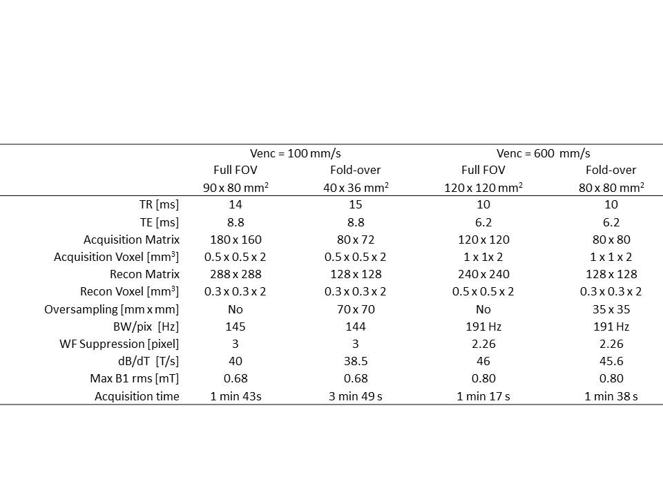
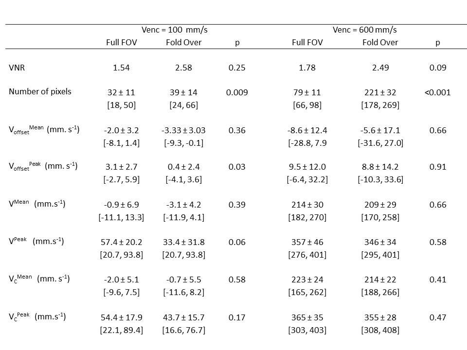
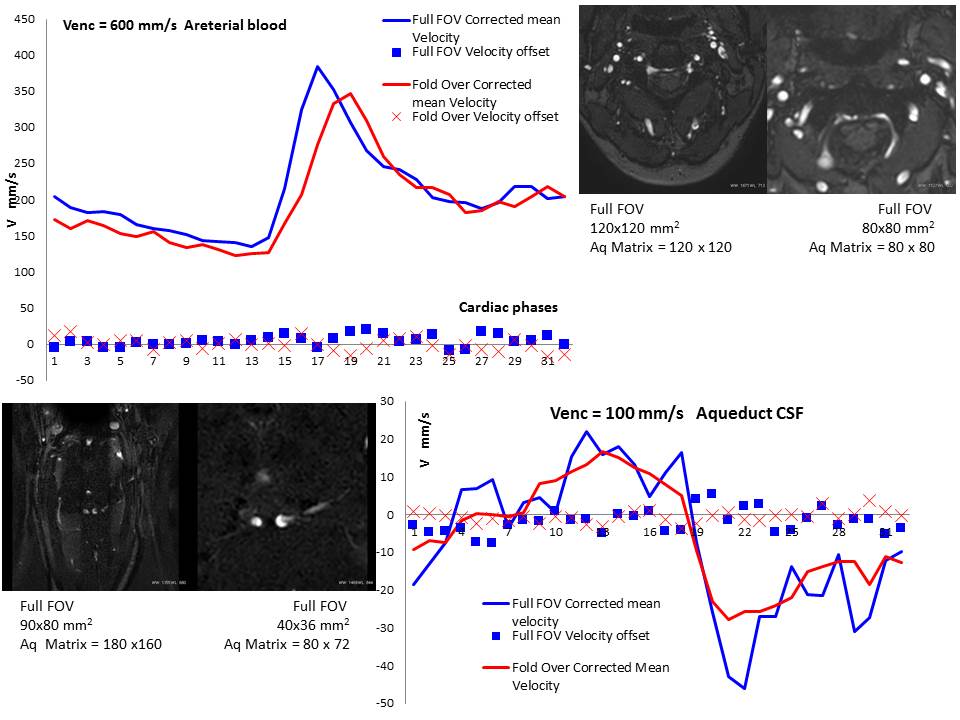
MRI Cartesian Fast Field Echo 2D Cine PC was performed on a 3T Achieva dStream scanner (Philips healthcare, The Bets, Netherlands) equipped with 32 channel head coil. The sequence was retrospectively gated over 32 heart phases = 32, Sense factor of 1.5, and a flip angle = 30°. Two through planes Venc values were chosen to encode the velocity of the cerebro-spinal fluid in the aqueduct (Venc = 100 mm/s) and the cervical blood flow in the arteries (Venc = 600 mm/s). Two schemes were applied for each Venc (Table 1). The 1st involves a full FOV to cover the entire object while the 2nd involves an oversampling to remedy for aliasing. Flow compensation was activated but the default background phase correction was switched off. Ten healthy controls adults (5 females) participated to the study. Following each in-vivo acquisition, a 5 liters container filled with water and solidified with agar gel to create a static phantom was positioned to replicate all of the sequences while respecting the same imaging parameters. Five variables were compared between the two schemes: the VNR and the velocity offset in the phantom (peak Voffsetpeak and mean Voffsetmean), the in-vivo peak (Vpeak) and mean velocity (Vmean) without phase-offset correction and with phase-offset correction (Vcpeak and mean Vcmean).Results
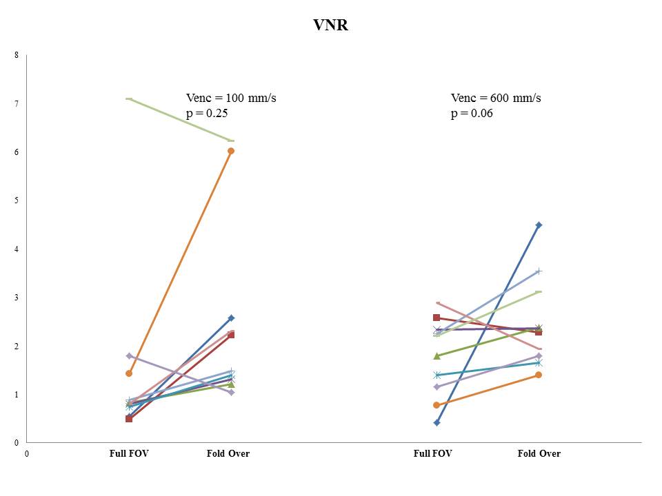
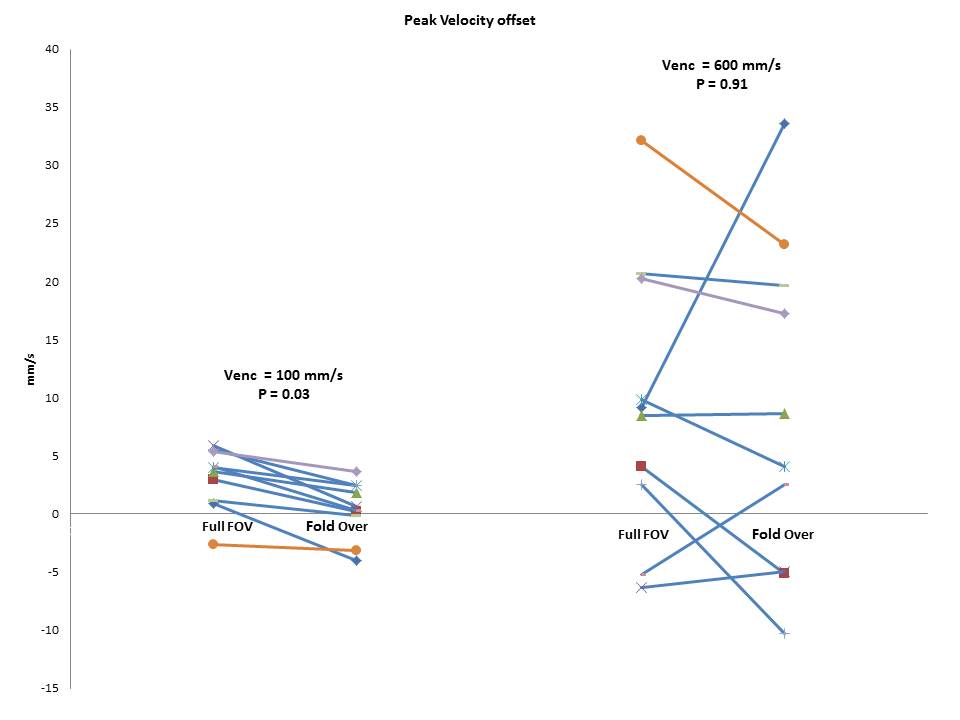
There is a significantly greater number of pixels with fold-over option in the CSF (p = 0.009) and blood flow (p < 0.001) compared to full FOV acquisition. The measured VNR on the static phantom for both velocity encodings (100 and 600 mm/s) did not differ between full FOV and fold-over option (Figure 1). The only other consequence of fold-over option is a significantly higher peak velocity offset (Voffsetpeak, p=0.03) compared to full FOV for Venc = 100 mm/s (Figure 2) but not for Venc = 600 mm/s (Figure 3). The mean velocity (Vmean) and peak velocity (Vpeak) measured in-vivo for both CSF and blood flows) prior to phase-offset background correction do not statistically differ (Table 2). Similarly, following velocity offset correction we did not observe any significant difference in Vcpeak and Vcmean (neither CSF flow nor blood flow).Discussion
The application of cerebral 2D Cine PC MRI has been proven to provide useful quantitative data for blood flow and CSF flow analysis. The results of our investigation show that the application of fold-over option to increase the number of pixels in the desired object of interest does affect neither the VNR nor the derived flow velocity variables, and can be used to acquire higher spatial resolution phase and magnitude images. The term φ(n) in the signal intensity is negligible and does not contribute to the reconstructed phase images with 2D cine PC-MRI.Acknowledgements
No acknowledgement found.References
1- Rüegger CM, Makki MI, Capel C, et al. An innovative approach to investigate the dynamics of the cerebrospinal fluid in the prepontine cistern: A feasibility study using spatial saturation-prepared cine PC-MRI. Eur J Radiol Open 2014 16(1):14-21
2- Capel C, Makki M, Gondry-Jouet C, et al. Insights into cerebrospinal fluid and cerebral blood flows in infants and young children. J Child Neurol. 2014 Dec;29(12):1608-15
3- Gatehouse PD, Rolf MP, Grave MJ, et al. Flow measurement by cardiovascular magnetic resonance: a multi-centre multi-vendor study of background phase offset errors that can compromise the accuracy of derived regurgitant or shunt flow measurements. J of Cardiovascular Magn Reson 2010, 12:5
4- Chang W, Loecher MW, Wu Y et al. Hemodynamic Changes in Patients with Arteriovenous Malformations Assessed Using High-Resolution 3D Radial Phase-Contrast MR Angiography. AJNR 2012 33:1565-72.
5- Macdonald J, Wieben O, Nagle S, Johnson K. Artifat reduction in 3D radial imaging with out-of-volume saturation pulse. Proceeding 24rd ISMRM (2016) 1831.
6- Stab Q, Wech T, Barth M. A data driven Nyquist ghost and gradient delay correction for navigator-free 3D planes on a paqddlewheel (POP) EPI. Proceeding 26th ISMRM (2677)
7- Bernstein MA, King EK, Zhou XJ. Chapters 8 and 11 in “Handbook of MRI pulse sequences”, ISBN 0-12-092861-2.
Figures