2958
Direct visualization the extracranial segments of the hypoglossal nerve on Enhanced 3D-SPACE-STIR sequence1Radiology, The First Affiliated Hospital of Xiamen University, Xiamen, China
Synopsis
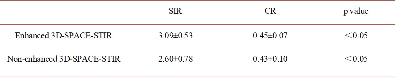
In our research, we compared enhanced and non-enhanced 3D-SPACE-STIR sequence in displaying the extracranial segments of the hypoglossal nerve. The SIR and CR of the nerve were higher on enhanced 3D-SPACE-STIR MR imaging than non-enhanced imaging (3.09±0.53 vs. 2.60±0.78 and 0.45±0.07 vs. 0.43±0.10, respectively, p<0.05). Enhanced 3D-SPACE-STIR sequence demostrates better background suppression than non-enhanced. The extracranial segments of the hypoglossal nerve (extracranial-carotid space and extracranial-anterior segments) can be traced continuously on non-enhanced 3D-SPACE-STIR sequence.
Introduction & purpose
The hypoglossal nerve distributes fibres to styloglossus, hyoglossus and genioglossus and to the intrinsic muscles of the tongue. If the nerve suffers either iatrogenic or pathological damage, the tongue, on protrusion, will deviate towards the affected side and there may also be wasting of the muscles on the affected side. The entire anatomical course the hypoglossal nerve include the medullary, cisternal, skull base, extracranial segments1. The first three segments are well depicted with segmental imaging approach such as CISS, DRIVE, FIASTA, SPGR, GRASS, MPRAGE or other MR sequences. While the extracranial segments (extracranial-carotid space and extracranial-anterior segments) are difficult to examined with MR or CT. However, it can be well examined with ultrasound2. Our research is to demostrating the extracranial segments of the hypoglossal nerve on Enhanced 3D-SPACE-STIR sequence.Materials and Methods
Eleven healthy volunteers (3 males and 8 females; age range, 25–62 years; mean age, 42±13 years) were enrolled and experienced non-enhanced and enhanced 3D-SPACE-STIR MR scan at 3.0 Tesla MRI (Ingenia Achieva TX, Philips Healthcare, Best, The Netherlands) using a 16-channel neurovascular head and neck coil (NV-16). Scanning parameters for 3D-SPACE-STIR sequence were, echo time (TE) = 410 msec, repetition time (TR) = 4800 msec, time of inversion (TI) = 245 msec, matrix=318×314, field of view (FOV) = 220 mm×220 mm, slice thickness = 1.2 mm, slice gap=-0.6mm, number of signal averages (NSA) = 2, scan time = 9.5 (min).We roughly divied the hypoglossal nerve into four segments to evaluated the MR images. Signal intensity of the nerve and lateral pterygoid muscle (LPM) were measured to calculate the signal intensity ratio (SIR) and contrast ratio (CR).Results
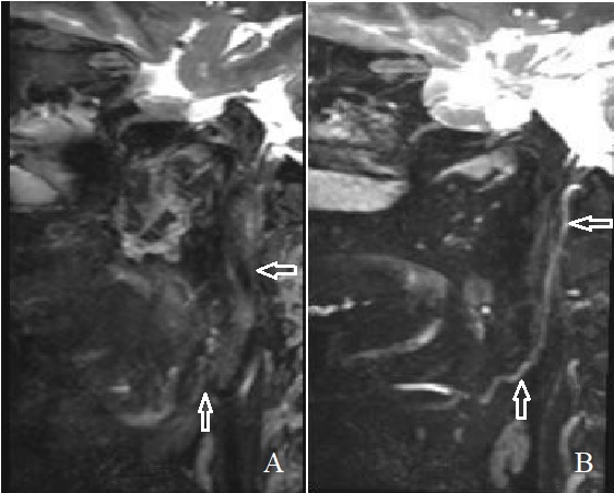
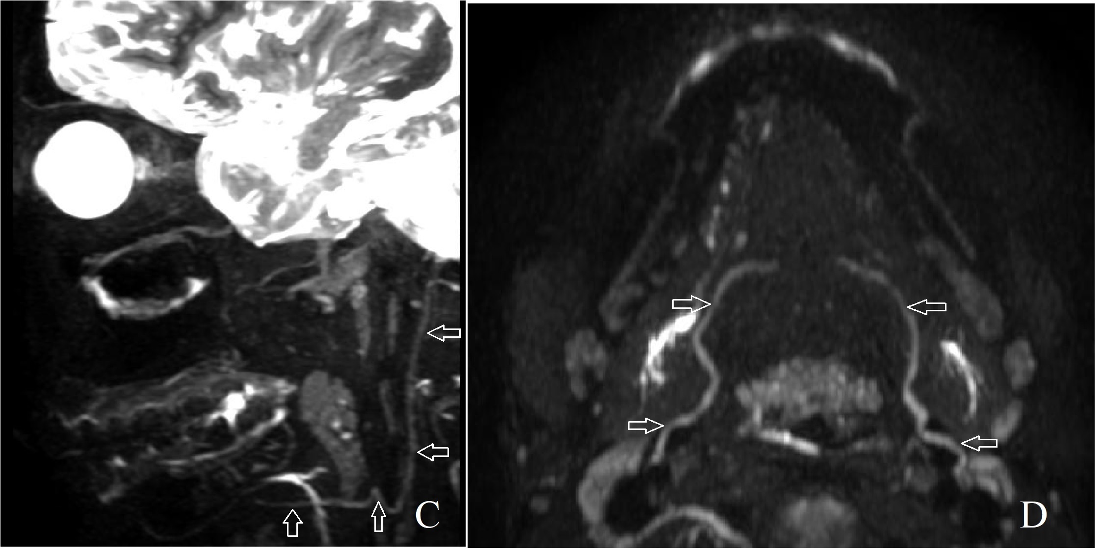
Enhanced 3D-SPACE-STIR sequence showed excellent direct visualization of the extracranial segments of the hypoglossal nerve than non-enhanced 3D-SPACE-STIR sequence (Figure 1). The extracranial segments (include the extracranial-carotid space and extracranial-anterior segments) of the hypoglossal nerve can be traced continuously (Figure 2). The SIR and CR of the nerve were higher on enhanced 3D-SPACE-STIR MR imaging than non-enhanced imaging (3.09±0.53 vs. 2.60±0.78 and 0.45±0.07 vs. 0.43±0.10, respectively, p<0.05) (Table 1).Discussion
The muscles, vessels, bones and lymphomas near the hypoglossal nerve demostrate hyperintensity mimick the nerve signal or even cover it on non-contrast 3D-SPACE-STIR imaging. However, these tissues turn to hypointensity after the injection of Gadolinium contrast agent due to a shorter transverse T2 relaxation time. The blood vessel barrier arround the hypoglossal nerve prevent the agent absorption so it maintain a intermediate signal. Therefore, the hypointensity background sets the relatively hyperintensity hypoglossal nerve off to advantage on enhanced 3D-SPACE-STIR imaging.Conclusion
Enhanced 3D-SPACE-STIR sequence demostrates better background suppression than non-enhanced. Extracranial segments of the hypoglossal nerve can be well depicted.Acknowledgements
No acknowledgement found.References
1. Alves P. Imaging the hypoglossal nerve[J]. European Journal of Radiology, 2010, 74(2):368-377.
2. Solomon M. Ultrasound of the Hypoglossal Nerve in the Neck: Visualization and Initial Clinical Experience with Patients[J]. Ajnr American Journal of Neuroradiology, 2016, 37(2):354.
Figures