2953
The influence of different MRI scan plane on radiomic features of the Nasopharyngeal Carcinoma1Cancer Hospital Chinese Academy of Medical Sciences, Shenzhen Center, shen zhen, China, 2GE Healthcare, bei jing, China, 3Huiying Medical Technology Co, bei jing, China
Synopsis
It is known that radiomics has drawn more attentions in radiological research, and has been applied to evaluate the prognosis of nasopharyngeal carcinoma (NPC). However, the influence of different MRI scan plane on the radiomic features of NPC has not been investigated. To address this issue, in current study, contrast-enhanced T1 weighted images (T1WI_C) in axial, coronal and sagittal views with and without axial T2 weighted images were applied to build the radiomics-based models for the prognosis of NPC. Our results showed that radiomics features derived from axial view of T1WI_C had the best performance for the prognosis prediction of NPC.
Introduction
Nasopharyngeal carcinoma (NPC) is a common malignant tumor in southeast China and southeast Asian countries [1]. Although patients with NPC have high 5 years of survival rate, there is still no effective means with regard to its prognostic analysis [2,3]. By extracting high throughput of quantitative descriptors from routinely acquired medical imaging data, recent advances in radiomics have provided insights in personalized medicine in oncologic practice related to tumor detection, strategy choice of treatment, prognosis analysis, and subtype classification. Some studies have reported that radiomics have great significance in prognosis prediction of NPC [4-6], while it only considered the radiomic features of T1WI images in axial view without adopting the other two orientation images. To address this issue, in current study, contrast-enhanced T1 weighted images (T1WI_C) in axial, coronal and sagittal views with and without axial T2 weighted images were applied to build the radiomics-based models for the prognosis of NPC.Materials and Methods
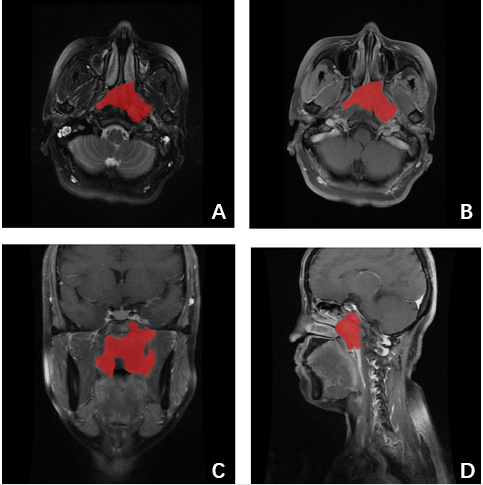
A total of 101 tissue biopsy confirmed patients of NPC were enrolled in this retrospective study. We chose the progression as the clinical endpoint. Patients were labeled as 1 if its progression-free survival (PFS) value smaller than 3 years, whereas the patients were labeled as 0 if its PFS value bigger than 3 years. All the patients were underwent with routing MR (Discovery MR750, GE, WI) including contrast-enhanced T1 weighted images with axial view, sagittal view, and coronal view (T1WI_AX_C,T1WI_SAG_C,T1WI_COR_C) and T2 weighted axial images (T2WI) from Dec 13th, 2012 to Dec 23th, 2015 in Cancer Hospital of the Chinese Academy of Medical Sciences. And these images were used to extract the radiomic features to analysis the PFS of NPC. Firstly, two radiologists with 3 years clinical experience were blinded to all patients’ information and manually outlined the ROI which enclose the boundary of tumor lesions on ITK-SNAP software. A representative set of patient images of NPC with outlined lesion regions that were used in feature derivations were shown in Fig.1. Then, all the original images and the ROI images were uploaded to Radcloud (http://radcloud.cn/, Huiying Medical Technology Co., Ltd, Beijing, China) to extract radiomic features. 1029 features were calculated and were classified as first order statistic, shape, gray level co-occurence matrix (GLCM), gray level run length matrix (GLRLM) and gray level size zone matrix (GLSZM), which were also transformed by exponential, square, square root, logarithm and wavelet (wavelet-LHL、wavelet-LHH、wavelet-HLL、wavelet-LLH、wavelet-HLH、wavelet-HHH、wavelet-HHL、wavelet-LLL). After that, the variance threshold and least absolute shrinkage and selection operator (LASSO) methods were used to select features that were most significantly associated with the progression on each sequence and the joint sequence respectively. And the selected features were used to construct radiomics-based models with three classifiers including logistic regression (LR), random forest (RF), and support vector machine (SVM) respectively. Finally, the predictive performance of those three classifiers were assessed with respect to the area under the curve (AUC) and the accuracy (ACC) to make the final model. The overall flow chart of the proposed method is illustrated in Fig.2.Results and Discussion
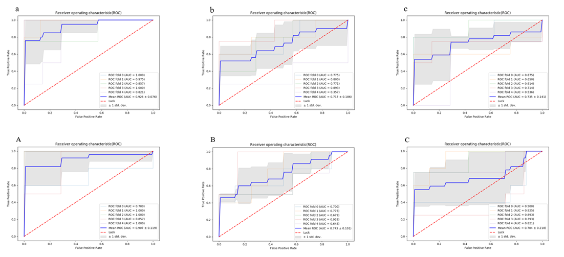
The Tab. 1 showed the prediction performance results of the three classifiers (LR, RF, and SVM) respectively on T1WI_C single sequences and the T1WI_C and T2WI joint sequences. From the table, we can see that the prediction performance on T1WI_AX_C sequence is better than others with LR (ACC=0.876, AUC=0.883), RF (ACC=0.845, AUC=0.760), SVM (ACC=0.930, AUC=0.926) in single sequences, and the prediction performance on T1WI_AX_C+T2WI is better than others with LR (ACC=0.951, AUC=0.941), RF (ACC=0.934, AUC=0.891), SVM (ACC=0.933, AUC=0.907) in joint sequences. Comparing the result of different classifier under different sequence, the prediction performance of SVM classifier is optimal to other classifiers. The ROC analysis of SVM is shown in Fig.3. T1WI_C images can reflect the tumor heterogeneity, T2WI images can reflect the cell density of tumor. The previous studies on radiomics feature analysis on T1WI_C images and T2WI images had achieved good results [5-6]. Our study shows the similar results that T1WI_C image and T2WI images can be used for the prognosis assessment of NPC, and the T1WI_C images shows the best performance.Conclusion
Our results showed that radiomics features derived from axial view of T1WI_C had the best performance for the prognosis prediction of NPC.Acknowledgements
No acknowledgement found.References
[1] Airoldi M, Gabriele P, Gabriele A M, et al. Induction chemotherapy with carboplatin and taxol followed by radiotherapy and concurrent weekly carboplatin taxol in locally advanced nasopharyngeal carcinoma.[J]. Cancer Chemotherapy & Pharmacology, 2011, 67(5):1027-1034.
[2] Fangzheng W, Chuner J, Quanquan S, et al. Addition of chemotherapy to intensity-modulated radiotherapy does not improve survival in stage II nasopharyngeal carcinoma patients [J]. Journal of Cancer, 2018, 9(11):2030.
[3] Huang PY, Cao KJ, Guo X, et al. A randomized trial of induction chemotherapy plus concurrent chemoradiotherapy versus induction chemotherapy plus radiotherapy for locoregionally advanced nasopharyngeal carcinoma [J].Oral Oncology. 2012, 48(10):1038-1044.
[4] Liu J, Mao Y, Li Z, et al. Use of texture analysis based on contrast-enhanced MRI to predict treatment response to chemoradiotherapy in nasopharyngeal carcinoma [J]. Journal of Magnetic Resonance Imaging Jmri, 2016, 44(2):445-455.
[5] Zhang S, Zhang B, Tian J, et al. Radiomics features of Multiparametric MRI as Novel Prognostic Factors in Advanced Nasopharyngeal Carcinoma.[J]. Clinical Cancer Research An Official Journal of the American Association for Cancer Research, 2017, 23(15):4259.
[6] Wang G, He L, Yuan C, et al. Pretreatment MR imaging radiomics signatures for response prediction to induction chemotherapy in patients with nasopharyngeal carcinoma[J]. European Journal of Radiology, 2018, 98:100-106.
Figures