2950
CAIPIRINHA-accelerated 3D constructive interference in the steady state MRI of trigeminal nerve: performance evaluation on image quality at 3.0TXiao-qing Sun1, Qiong Zhang2, Xiao-yue Zhou3, Qing-lei Shi4, and Wei Wang1
1China-Japan Union Hospital of Jilin University, Changchun, China, 2Siemens Shenzhen Magnetic Resonance Ltd, Shanghai, China, 3MR Collaboration, Siemens Healthcare Ltd, Shanghai, China, 4MR Scientific Marketing, Diagnostic Imaging, Siemens Healthcare Ltd, Beijing, China
Synopsis
This study aimed to compare controlled aliasing in parallel imaging results in higher acceleration (CAIPIRINHA) accelerated 3D constructive interference in the steady state (3D-CISS) with conventional CISS, whether image quality can be achieved under the condition of shortening scanning time,we evaluated imaging quality of the trigeminal nerve in 14 healthy subjects,no significant differences of SNR and CNR were found between the two sequences, in other words, CAIPIRINHA- accelerated 3D CISS can improve scanning efficiency without affecting image quality.
Objective
To evaluate the imaging quality of the trigeminal nerve between controlled aliasing in parallel imaging results in higher acceleration (CAIPIRINHA) accelerated 3D constructive interference in the steady state (3D-CISS) and conventional CISS that without acceleration factors under the same imaging parameters at 3.0T.Methods
Fourteen normal volunteers (female: male: 6: 8, mean age: 24 ±1.04) were examined with 3D-CISS and CAPIRINHA- accelerated CISS sequences at 3.0 T scanner (MAGNETOM Spectra, Siemens AG, Erlangen, Germany). The scan parameters are shown in table 1. In order to quantitative analysis the image quality of CAIPIRINHA- accelerated CISS sequence and traditional 3D CISS sequence, two radiologists with 10 years working experience in neurology measured the signal intensity of trigeminal nerve and pons, and calculated the signal noise ratio (SNR) and the contrast to noise ratio (CNR) of the trigeminal nerve,and then the subjective score of images on trigeminal nerve details displaying, the quality of peripheral anatomy and the degree of artifact based on CAIPIRINHA- accelerated CISS and 3D-CISS sequence were evaluated by the same two radiologists according to a three-point method (1: anatomical structure can be shown, but the image is blurry and cannot be used for diagnosis; 2: the anatomic structure is partly fuzzy but the image can be used for diagnosis; 3: the edge of anatomic structure is shown clearly, no artifact). The agreement of two readers was evaluated by Kappa test (k<0.20, poor; 0.21–0.40, fair; 0.41–0.60,moderate; 0.61–0.80, good; and 0.81–1.00, excellent).All the quantitative indexes were tested by paired-samples t test and the qualitative indexes were tested by Wilcoxon signed ranks test using SPSS software 18.0 (SPSS, Chicago, IL ), P value<0.05 was considered statistically significant difference.Results
No significant differences of SNR was found between traditional 3D-CISS and CAIPIRINHA- accelerated 3D CISS [(66.32±20.91) vs (59.81±18.80),P=0.118];The differences of CNR were also not significant between traditional 3D-CISS and CAIPIRINHA- accelerated 3D CISS [(8.29±15.29) vs (11.53±10.71),P=0.814]. The subjective evaluation scores of both readers didn’t show significant differences (table 2). Interobserver agreement of image quality has good consistency (K=0.80).Conclusion
Compared to conventional 3D-CISS, CAIPIRINHA- accelerated CISS sequence can significantly shorten the scanning time without affecting the image quality under the same scanning parameters at 3.0T.Discussion
With the development of MR technology, it is of great help to diagnose the etiology of trigeminal neuralgia caused by vascular compression. Most of the patients with trigeminal neuralgia cannot insist on long scanning, and the weak exercise of the patient can significantly reduce the image quality. This requires that the scanning time be shortened without affecting the image quality in the scanning process. The recently emerging CAIPIRINHA- accelerated 3D CISS as an optimized parallel acquisition technique, can significantly decrease scanning time under the premise of guaranteeing the image quality.Acknowledgements
No acknowledgement found.References
1) Fritz J, Fritz B, Thawait GG, Meyer H, Gilson WD, Raithel E (2016) Three-dimensional CAIPIRINHA SPACE TSE for 5-minute high-resolution MRI of the knee. Invest Radiol 51(10):609–6172) Kalia V, Fritz B, Johnson R, Gilson WD, Raithel E, Fritz J (2017)CAIPIRINHA accelerated SPACE enables 10-min isotropic 3D TSE MRI of the ankle for optimized visualization of curved and oblique ligaments and tendons. Eur Radiol 27(9):3652–3661Figures

Figure 1 Comparison of (A,C) CISS-3D and(B,D) CAIPIRINA- accelerated 3D CISS images in a 24-year-old male with no trigeminal lesion. There is no significant difference between the two sequences in demonstrating trigeminal nerve.
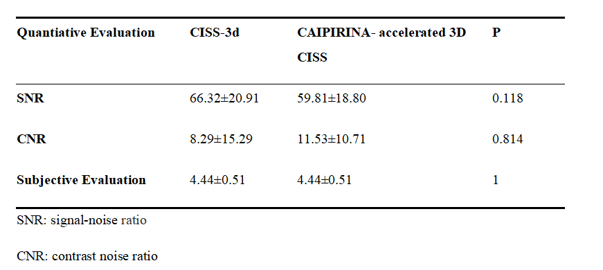
Table 2 Comparison of quantitative index for image quality and subjective scores between CISS-3D and CAIPIRINA- accelerated CISS
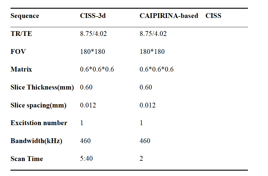
Table1 MRI scanning parameters of CAIPIRINHA- accelerated CISS and conventional CISS sequence.