2947
Preliminary Results of 31P Magnetic Resonance Spectroscopy for Human Submandibular and Parotid Glands1Department of Diagnostic Imaging and Nuclear Medicine, Graduate School of Medicine, Kyoto University, Kyoto, Japan
Synopsis
We previously performed 31P MRS of the human parotid gland,
but no such study of the submandibular gland has yet been reported. The present work was
designed to evaluate the potential of 31P MRS, using 3D CSI, to simultaneously
measure the phosphorus metabolites of human submandibular and parotid glands. Three
healthy volunteers were examined before and after oral intake of vitamin C. Corresponding
spectra revealed intense adenosine triphosphate (ATP) and phosphocreatine peaks.
Following vitamin C intake, β-ATP decreased in both glands. This preliminary finding indicates that 31P
MRS could provide unique information on the bioenergetics of submandibular and
parotid glands.
INTRODUCTION
The major salivary glands are the paired parotid, submandibular, and sublingual glands. While stimulated saliva production is largely (60–70% of total) derived from the parotid glands and balanced by other glands, resting (unstimulated) saliva production occurs primarily from the submandibular and sublingual glands.1 We have previously reported 31P magnetic resonance spectroscopy (MRS) data of the human parotid gland2; however, to the best of our knowledge, no similar study of the human submandibular gland has yet been reported. The present work was designed to evaluate the potential of 31P MRS to measure the phosphorus metabolites of human submandibular and parotid glands.METHODS
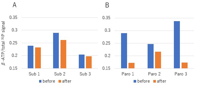
Subjects and design: Three healthy volunteers (age 33–52 y) were examined before and after intake of tablets containing approximately 200 mg of vitamin C.
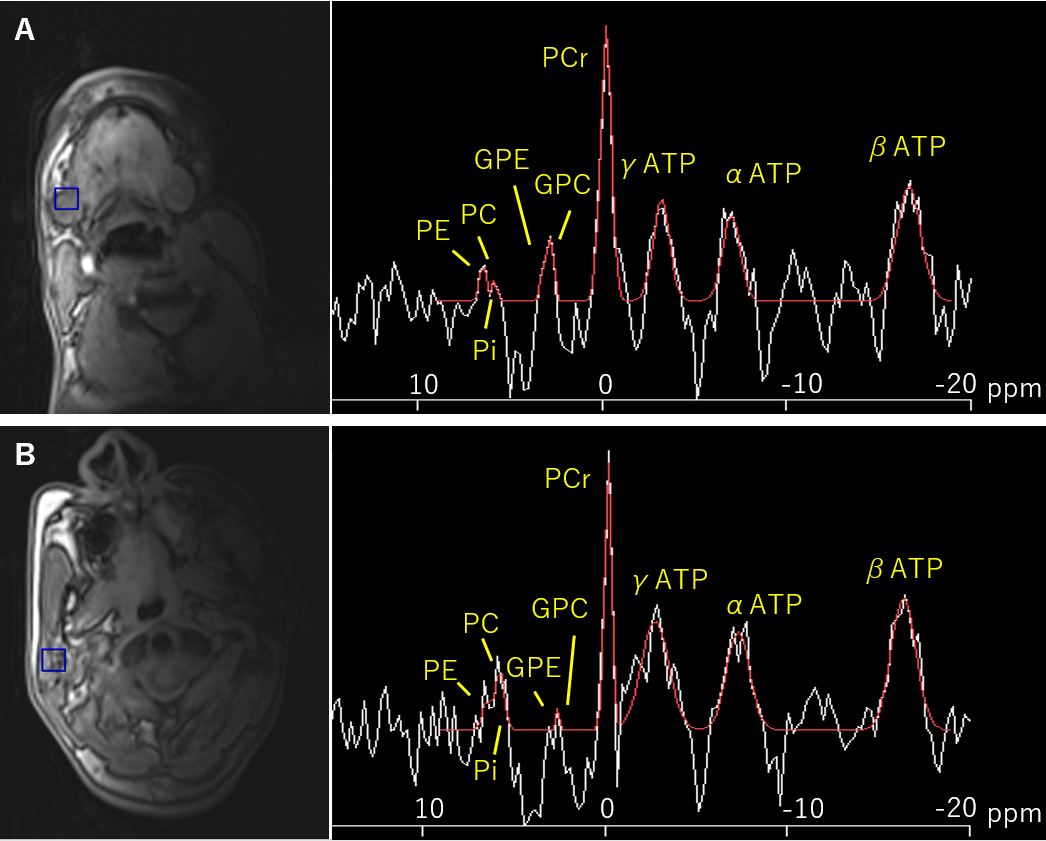
MR experiment: Subjects were placed in a right lateral position in the magnet. T1 weighted ultrafast gradient echo images were collected with 4.5-mm thick slices covering the right parotid and submandibular glands. All 3D 31P spectroscopic imaging data were acquired on a 3.0 T MRI (MAGNETOM Skyra, Siemens Healthcare, Erlangen, Germany) using a 31P/1H loop transmit-and-receive RF coil (Takashima Seisakusho Co. Ltd., Tokyo, Japan) with a diameter of 140 mm and a 3D CSI-FID pulse sequence (TR/TE: 1000 ms/2.3 ms, bandwidth: 3000 Hz, 128 FID data points/zero-filling to 256). Proton-decoupling during excitation and nuclear Overhauser effect were used to improve the spectral resolution. A 200 mm x 200 mm x 200 mm field of view (FOV) was phase encoded to 16 x 16 x 16 matrix, resulting in a nominal voxel size of 2.2 cm x 2.2 cm x 2.5 cm or 12.3 cm3. Weighted averaging (12 averages) led to a total acquisition time of about 10 min 32 s.
Data analysis: All spectra were analyzed with Syngo software (Syngo Spectroscopy Evaluation, Siemens Medical Systems, Erlangen, Germany). The following metabolites were fitted: phosphocholine (PC), phosphoethanolamine (PE), inorganic phosphate (Pi), glycerophosphoethanolamine (GPE), glycerophosphocholine (GPC), phosphocreatine (PCr), and adenosine triphosphate (ATP).
RESULTS
Clear 31P spectra were obtained for all volunteers. The voxel was set such that signals from other areas were excluded. The spectra revealed large ATP and PCr intensities in both submandibular and parotid glands (Fig. 1). β-ATP resonance over the total phosphorus signal decreased after the intake of vitamin C tablets; the decrease was less in the submandibular glands than in the parotid glands (Fig. 2).DISCUSSION
This is the first in-vivo study to investigate the major phosphorus metabolites of human submandibular gland using 3D CSI. Our results show that the proposed 31P MRS technique could simultaneously record the spectra of submandibular and parotid glands with acceptable quality and measurement time. The PCr signal in submandibular and parotid glands indicates a high and rapid energy demand in the organ. This result is consistent with data from our earlier study2 and previous animal models.3,4 Furthermore, continuous imaging with vitamin C intake showed that it may even be possible to capture β-ATP changes during saliva secretion. Decrease of β-ATP in the submandibular gland was less than that in the parotid gland, which may be related to the difference in saliva production.CONCLUSION
This preliminary study showed that 31P MRS may provide unique information on the bioenergetics of human submandibular and parotid glands.Acknowledgements
No acknowledgement found.References
1. Dawes, C. and C. M. Wood. The contribution of oral minor mucous gland secretions to the volume of whole saliva in man. Arch Oral Biol. 1973;18(3): 337-342.
2. Sato T., Isoda H., Arizono S., et al. In vivo 31P magnetic resonance spectroscopy of human parotid gland. Proceedings 26th ISMRM, Paris, 2018.
3. Seo, Y., Steward, M. C., Mackenzie, I. S., et al. Acetylcholine-induced metabolic changes in the perfused rabbit mandibular salivary gland studied by 31P-NMR spectroscopy. Biochim Biophys Acta. 1988;971(3): 289-297.
4. Murakami, M., Seo, Y., Watari, H., et al. Dissociation of fluid secretion and energy supply in rat mandibular gland by high dose of ACh. Am J Physiol. 1988;254(5 Pt 1): G781-787.
Figures