2943
Clinical validation of synthetic MRI in assessing composition of Carotid Atherosclerotic plaques: initial experience1MR Application China, GE Healthcare, Shen Yang, China, 2Radiology Department of China Medical University First Hospital, Shenyang, China, 3GE Healthcare, MR Application China, Beijing, China, 4GE Healthcare China, Beijing, China
Synopsis
The current study aims to determine the accuracy of synthetic MRI sequences generated from post-acquisition processing of a single sequence compared with sequences imaged from conventional multi-contrast Weighted (MCW) methods in assessing the composition of Carotid artery plaques like calcification and intraplaque hemorrhage (IPH). It was concluded that synthetic MRI sequences are comparable to conventional MCW methods.
Introduction
Vulnerable plaques may lead to cerebral ischemic events, the composition of the plaque play an important role in its vulnerability 1. Multi-contrast MRI including TOF, T1w, T2w and PD has been used to identify the plaque composition, given that different contents have different features in different image contrasts 2. In convention, different images contrasts are acquired separately that may give rise to several practical issues: long scan time, patient motion, image spatial mis-registration. These practical issues not only render the widespread of the MR carotid plaque imaging, also may introduce errors in the tissue composition. Synthetic MR is a novel imaging method that may offer simultaneous image contrasts in a single scan, which may elevate the practical issues. In this work, we investigate the utility of the multiple-contrast generated in synthetic MR and perform a non-inferiorty test.Methods
After providing informed consent, seven patients who were scheduled for (Carotid endarterectomy) CEA were enrolled in this study , ethical approval and consent forms were obtained. All the patients underwent MR scans on a 3.0T whole body scanner (GE Signa Pioneer, WI), and the scan protocol consisted of both conventional multi-contrast MR and synthetic MR. Matching spatial resolution of 0.7x0.7x3 mm3 were used in conventional and synthetic MR scans. In conventional MR, PD, T1w and T2w 3D black blood FSE were scanned as well as the TOF. Two experienced radiologist reviewed the conventional MR images and synthetic MR images, and independently identified different compositions according to the criteria in 3,4: IPH, noIPH and Calcification. The areas of identified hemorrhage and calcification regions obtained using conventional and synthetic MR are respectively measured, SPSS19.0 was used to analyze the data. Student t-test was performed to assess the agreement, P < 0.05 was considered statistically significant.Result
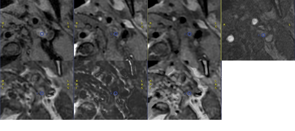
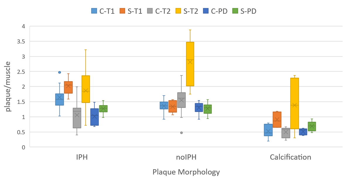
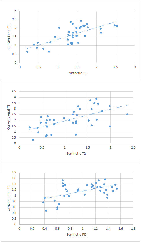
A total of 140 image locations were acquired from all 7 patient exams. Using the criteria 2, IPH was identified at 20 image locations and calcification at 7 image locations. Figure.1 illustrate matching location of conventional MCW and synthetic MRI images : the IPH regions can be identified at both conventional MCW images and synthetic MR images. The box diagram in figure 2 compares diagnostic efficacy between conventional MRI and synthetic MRI(Figure.2). For all the locations identified, strong correlations were observed between MCW (T1,T2,PD) and synthetic MRI(T1, T2, PD). However, statistical calculations show that the values measured by synthetic T1 are upper than conventional T1 (on an average of 18.7%), synthetic T2 are upper than conventional T2 (on an average of 86.9%), synthetic PD are upper than conventional PD (on an average of 15.1 %), as is shown in Figure.3.Discussion
Compared to conventional multi-contrast MRI, synthetic MR has the intrinsic advantages of shortened overall scan time, perfect registration among different image contrasts. It has great potential in identifying plaque compositions. In this work, the equivalency of synthetic MR in performing composition identification is tested using a non-interiority test with conventional MR. Excellent agreements on the compositions identified by the two methods were observed, which support the use of synthetic MR in this application. Further investigations on other clinically valuable compositions such as necrotic core and fiber cap may be performed to verify synthetic MR’s performance.Conclusion
The multiple contrasts offered by synthetic MR may be used for identifying the carotid plaque composition.Acknowledgements
No acknowledgement found.References
[1] Naghavi M, Libby P, Falk E, et al. From vulnerable plaque to vulnerable patient: a call for new definitions and risk assessment strategies: part I. Circulation 2003;108:1664–1672.
[2] den Hartog AG, Bovens SM, Koning W, Hendrikse J, Luijten PR, Moll FL, Pasterkamp G, de Borst GJ. Current status of clinical magnetic resonance imaging for plaque characterisation in patients with carotid artery stenosis. Eur J Vasc Endovasc Surg 2013;45:7-21.
[3] OtaH, YuW, UnderhillHR, Oikawa M,Dong L, Zhao X, etal. Hemorrhage andlarge lipid-rich necrotic cores are independently associated with thin or ruptured fibrous caps: an in vivo 3T MRI study. Arterioscler Thromb Vasc Biol 2009;29:1696–701.
[4] Yuan C, Mitsumori L, Beach K, Maravilla K. Carotid atherosclerotic plaque: noninvasive MR characterization and identification of vulnerable lesions. Radiology 2001;221:285–99.
Figures