2936
Skin blood flow functions as a potential proxy for cerebral blood flow in adults with sickle cell disease1Department of Biomedical Engineering, Wayne State University, Detroit, MI, United States, 2Department of Radiology, Wayne State University, Detroit, MI, United States, 3Biogen Inc., Cambridge, MA, United States, 4Department of Hematology-Oncology, Wayne State University, Detroit, MI, United States, 5MR Innovations, Detroit, MI, United States, 6Centre for Human Drug Research, Leiden, Netherlands, 7Bioverativ, a Sanofi company, Waltham, MA, United States, 8United Neuroscience, Hauppauge, NY, United States
Synopsis
Even though the prevalence of stroke in adults with sickle cell disease (SCD) is higher compared to children, no accepted screening measures are available for identifying adults with SCD at high risk for strokes. Despite cerebral blood flow (CBF) from transcranial Doppler being an established surrogate measure of stroke risk in children, it is not feasible in adults and MRI is uneconomical and inefficient for screening. We examined skin blood flow using laser speckle contrast imaging as a peripheral surrogate of CBF using MRI. Skin blood flow was highly correlated with CBF in sickle cell patients with excellent test-retest reliability.
Introduction
Cerebrovascular accidents (CVA), including stroke, are one of the major causes of morbidity and mortality in patients with sickle cell disease (SCD) resulting from vaso-occlusion of major cerebral arteries. Experience with transcranial Doppler (TCD) ultrasonography in children highlights the importance of cerebral blood flow (CBF) assessment in optimal management of CVA risk1,2. While the rates of CVA are even higher in adult SCD populations3 TCD has not proven to be as valuable a measure in the adult SCD population and less information is available about CBF in adults with SCD4. This is likely due to closure of ultrasound windows in adults and a more complex pathophysiology of stroke in adults with SCD4. CBF has been an established surrogate measure of stroke risk in children. Despite magnetic resonance imaging (MRI) being an optimal imaging technique to evaluate CBF, the cost, time and inconvenience of MRI scans makes it uneconomical and inefficient for the routine screening and surveillance of SCD patients for stroke risk. Therefore, to explore more pragmatic surrogate biomarkers of stroke in SCD patients, in this study we investigate skin blood flow using laser speckle contrast imaging (LSCI) as a potential surrogate biomarker of altered CBF in adults with SCD using MRI.Materials and Methods
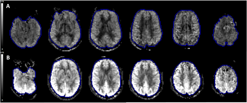
We enrolled SCD patients (N=18 at imaging visit 1 with 1 subject lost to follow-up) not receiving routine blood transfusion protocols and matched healthy volunteer controls (N=6). All MRI scans were performed at 3.0 Tesla (Siemens Healthineers, Erlangen, Germany). We investigated CBF in adults with SCD using 2 MRI techniques: arterial spin labeling (ASL) (Figure-1) and phase contrast (PC-MRI) (Figure-2). Additionally, we measured skin blood flow using LSCI by using perfusion and re-perfusion criteria (Figure-3). Imaging data was acquired in 2 visits for both MRI and LSCI. The association between the CBF and skin blood flow was investigated using the Pearson’s correlation coefficient. The intraclass correlation coefficient (ICC) was estimated for assessing test-retest reliability of the CBF and skin blood flow.Results
A strong correlation was found between the CBF quantified using ASL (Figure-4) or PC-MRI (Figure-5) and the baseline blood flow in the skin as measured using LSCI in the subjects with SCD (ASL vs. LSCI: (r = 0.56; p=0.016); PC-MRI vs. LSCI: r = 0.67, p=0.002)). Figures 4 and 5 also show that this association was found to be significant and consistent during imaging visit 2 (ASL vs. LSCI: (r = 0.51; p=0.038); PC-MRI vs. LSCI: r = 0.63, p=0.006).Discussion
Baseline blood flow in the skin as measured using LSCI was found to be a strong predictor of the average CBF quantified using ASL or PC-MRI in adult subjects with SCD. In addition, this correlation was highly reproducible in test-retest process. The change in rheological properties of sickled RBCs and altered vaso-motor function may affect the hemodynamic properties of the CNS and peripheral system proportionally, which is captured in the strong and reproducible correlation between CBF and skin blood flow in SCD patients. LSCI might be an inexpensive and clinically feasible technique to indirectly assess hemodynamic changes in the brain. With additional independent validation, LSCI may have a favorable cost:benefit ratio based on these data, and implementation of brain MRI and skin LSCI as exploratory endpoints to evaluate clinical correlates of increased blood flow in SCD clinical trials is justified.Conclusion
Good correlation was found between alterations in CBF and skin blood flow in adult SCD patients with significant longitudinal stability suggesting that skin blood flow measurement using LSCI may be a more convenient proxy than MRI for routine hemodynamic evaluation of this population.Acknowledgements
No acknowledgement found.References
1. Adams, R., et al., Long‐term stroke risk in children with sickle cell disease screened with transcranial Doppler. Annals of neurology, 1997. 42(5): p. 699-704.
2. Strouse, J.J., et al., Inverse correlation between cerebral blood flow measured by continuous arterial spin-labeling (CASL) MRI and neurocognitive function in children with sickle cell anemia (SCA). Blood, 2006. 108(1): p. 379-381.
3. Jordan, L.C. and M.R. DeBaun, Cerebral hemodynamic assessment and neuroimaging across the lifespan in sickle cell disease. Journal of Cerebral Blood Flow & Metabolism, 2017: p. 0271678X17701763.
4. Jordan, L.C., et al., Non-invasive imaging of oxygen extraction fraction in adults with sickle cell anaemia. Brain, 2016. 139(3): p. 738-750.
Figures