2932
Parallel Imaging Hadamard Encoded pCASL with Efficient Use of Proton Density Weighted Image1MR Applications and Workflow, GE Healthcare, Tokyo, Japan, 2Radiological Center, University of Fukui Hospital, Fukui, Japan, 3MR Application & Workflow, GE Healthcare, Calgary, AL, Canada, 4Department of Radiology, University of Fukui, Fukui, Japan
Synopsis
A different method of parallel imaging acceleration extracting coil sensitivity maps necessary for using CG SENSE from a proton density weighted image was proposed. Single and multiple post labeling delay (PLD) scan were accelerated while maintaining image quality. The proposed technique is expected to achieve both the robustness of CBF quantification and short imaging time that can be applied clinically.
Purpose
pCASL of 3D Fast Spin-Echo with stack-of spirals (SOSP) uses an efficient data collection and is useful for cerebrovascular disorders and brain tumors with clinically available imaging time. In principle, since there is flow velocity dependence, arterial transit delay needs to be compensated for using a multi-delay time point measurement to increase the robustness of cerebral blood flow quantification1. Although it is important to achieve both robustness and imaging time that can be applied clinically, additional scanning from a single time point measurement requires extension of imaging time even with Hadamard encoded acquisition in the multiple time delay scan2. In ASL, the recent work of parallel imaging applied to SOSP has been performed with SPIRIT and 1 D acceleration in the slice direction3,4. In this work, we propose a different method of parallel imaging acceleration extracting coil sensitivity maps necessary for using CG SENSE from proton density weighted image. It is beneficial that the additional external sensitivity reference and the gradient waveform modification of spiral trajectory for densely fulling the central k-space portion are not required.Methods
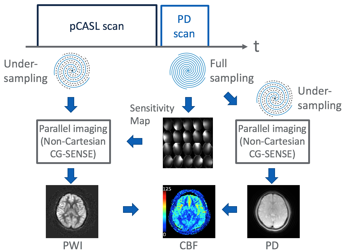
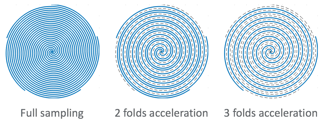
A schematic diagram is shown in Fig.1. The ASL scan used for perfusion weighted imaging (PWI) and the proton density weighted scan for calculating quantify cerebral blood flow are performed. The ASL acquisition is undersampled using fewer spiral interleaves than the Nyquist criteria. Two folds acceleration means only 1/2 of the interleaves (Fig.2). All the slice encodings in one spiral interleave are acquired. The proton density weighted image is fully sampled to extract coil sensitivity maps and is undersampled from the full sampled data after acquisition followed by parallel imaging reconstruction to calculate CBF. The coil sensitivity maps are generated from all the coil images with low resolution5. Reconstruction is performed using the acquired k-space data and conjugate Gradient Non-Cartesian SENSE (CG-SENSE) algorithm6 which employs the non-uniform fast Fourier transform (NUFFT)7-8 for gridding. Experiments were conducted on a 3.0T Scanner (Discovery MR 750, GE Healthcare, Waukesha, WI, U.S.A.) with 32 channel head coil. It was compared with data obtained by healthy volunteer scans under IRB approval. The ASL scan obtained a single and seven multiple PLD perfusion map. Hadamard time encoding was used in the multiple time point PLD scan. Background suppression was used with 4 TIs to suppress the background static tissues9. Vessel suppression was not used. Spatial resolution 3.72 mm x 3.72 mm with number of arms 8, slice thickness 4 mm, NEX 1, label duration 3.5 sec, PLD 700 ms for single delay and 700, 1200, 1700, 2200, 2700, 3200 and 3700 ms for seven delay. A long-labeling acquisition with labeling or control for the entire 3500 ms preparation period was also acquired to generate CBF map.Results and Discussions
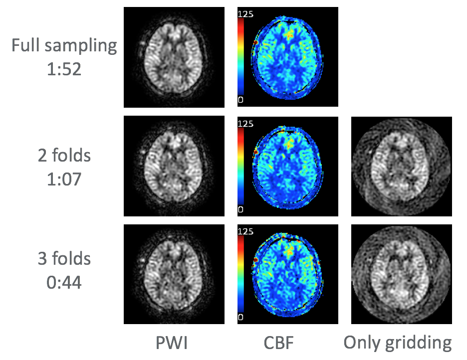
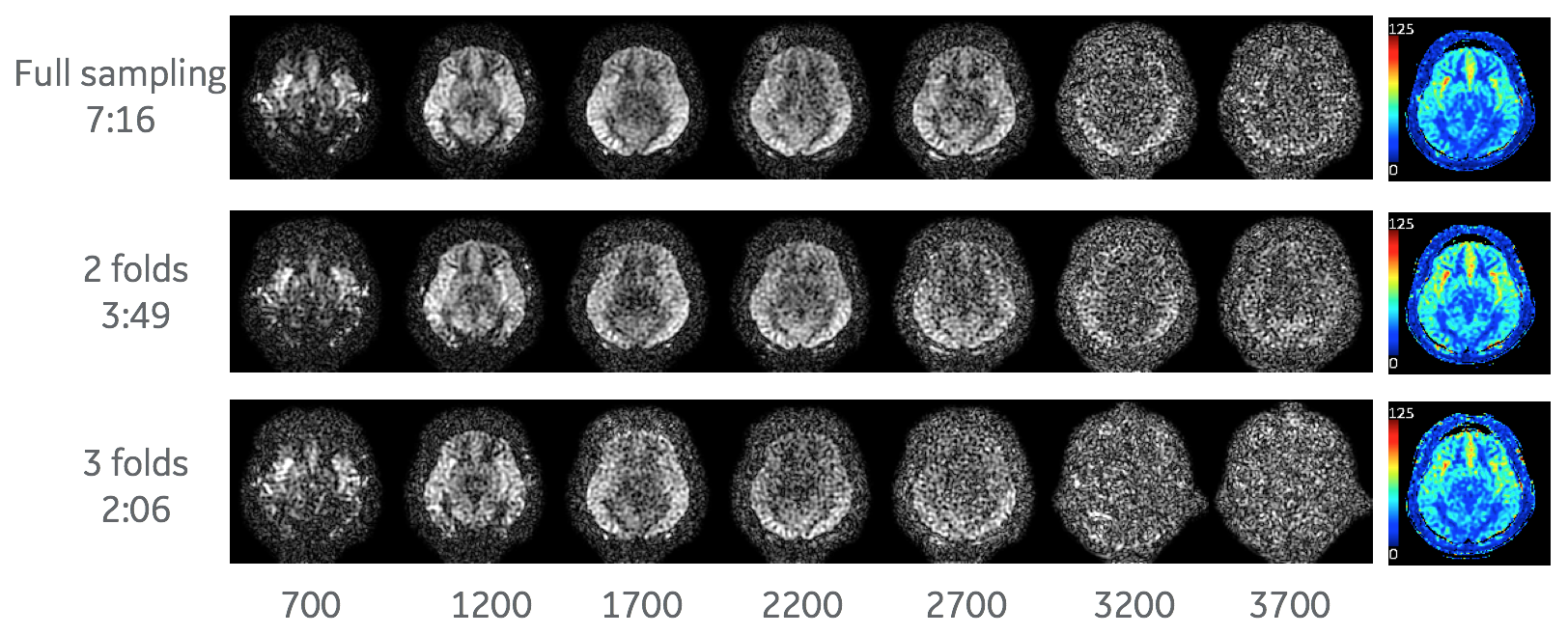
Fig.3 and Fig.4 show the result of single and seven PLD scans, respectively. The perfusion-weighted images were obtained without significant artifact. Compared to the full sampling scan, the CBF maps were similar for all acceleration scan while the PWI signal intensity decreased in proportion to higher folds scan. For the multiple PLD scans, the PWI at each time points suffered from low SNR because the arterial spin labeling preparation of 3500 ms was divided into 500 ms, resulting in being low perfusion signal and also undersampled data were reconstructed by parallel imaging. The similar tendency was found in a separate experiment that full sampling data were retrospectively undersampled to simulate SOSP undersampling, which is not shown in the abstract.Conclusion
We demonstrated that multiple-delay encoded scan was accelerated by generating coil sensitivity maps from a proton density weighted image enabling parallel imaging technique while maintaining image quality. The CBF maps was similar to the maps obtained by full sampling even at the imaging time that was speeded up to three folds. As contribution to perfusion from labeled blood in ASL measurements is as low as a few percentage, clinical evaluation is needed for the concern of SNR deterioration in combination with parallel imaging.Acknowledgements
No acknowledgement found.References
1. Guo J, Holdsworth SJ, Fan AP, Lebel MR, Zun Z, Shankaranarayanan A, Zaharchuk G. Comparing accuracy and reproducibility of sequential and Hadamard-encodedmultidelay pseudocontinuous arterial spin labeling for measuring cerebral blood flow and arterial transit time in healthy subjects: A simulation and in vivo study. J Magn Reson Imaging. 2018 Apr;47(4):1119-1132.
2. Gunther M. Encoded continuous arterial spin labeling. In: ISMRM workshop on cerebral perfusion and brain function: Novel techniques and applications, Salvador da Bahia, Brazil, 28 July–1 August 2007, abstract number: 3.
3. Chang YV, Vidorreta M, Wang Z, Detre JA. 3D-accelerated, stack-of-spiralsacquisitions and reconstruction of arterial spin labeling MRI. Magn Reson Med.2017 Oct;78(4):1405-1419.
4. Vidorreta M, Wang Z, Chang YV, Wolk DA, Fernández-Seara MA, Detre JA.Whole-brain background-suppressed pCASL MRI with 1D-accelerated 3D RAREStack-Of-Spirals readout. PLoS One. 2017 Aug 24;12(8).
5. McKenzie CA, Yeh EN, Ohliger MA, Price MD, Sodickson DK. Self- calibrating parallel imaging with automatic coil sensitivity extraction. Magn Reson Med 2002;47:529–538.
6. Pruessmann KP, Weiger M, Bornert P, Boesiger P. Advances in sensi- tivity encoding with arbitrary k-space trajectories. Magn Reson Med 2001;46:638–651.
7. Fessler JA, Sutton BP. Nonuniform fast Fourier transforms using min- max interpolation. IEEE Signal Process 2003;51:560–574.
8. Jeffrey A Fessler, "Michigan Image Reconstruction Toolbox".
9. Dai W, Shankaranarayanan A and Alsop DC. Volumetric measurement of perfusion and arterial transit delay using hadamard encoded continuous arterial spin labeling. Magn Reson Med 2013; 69: 1014–1022.
Figures