2928
Multiple overlapping slab 4D-flow imaging using distributed-spiral acquisition with magnetization transfer (MT) preparation for inflow signal enhancement and simultaneous 3D time-of-flight (3D-TOF) angiogram1Department of Physics, University of Wisconsin - Madison, Madison, WI, United States, 2Department of Medical Physics, University of Wisconsin - Madison, Madison, WI, United States, 3Department of Radiology, University of Wisconsin - Madison, Madison, WI, United States
Synopsis
Quantification of 4D-flow MRI relies on signal magnitude and phase of blood but can be affected by high signal intensities from surrounding tissues. By acquiring imaging volume in multiple overlapping slabs, together with MT preparation further to saturate tissue, 3D-TOF inflow enhancement can be used to improve vessel signal in the 4D-flow magnitude image. The scan-time penalty associated with acquiring multiple slabs was avoided by accelerating the scan with undersampled distributed-spiral acquisition. The proposed method greatly increased vessel contrast and improved depiction of blood vessels, which for 4D-flow MRI translate to more reliable flow quantification and vessel segmentations.
Introduction
By encoding velocities in the phase of the signal, 4D-flow MRI enables direct visualization of blood flow and quantitative hemodynamic analysis. However, the precision and accuracy of quantified parameters depends on the signal magnitude, quality of vessel segmentation, and partial voluming effects from nearby background signal. Using an angiogram from 3D-time-of-flight (TOF) can provide an alternative means of segmentation but does not improve the underlying velocity data. In principle, TOF effects only influence signal magnitudes and this mechanism may be combined with 4D-flow MRI to enhance vessel signal while suppressing background tissue. However, multiple overlapping slabs required are required for maximal 3D-TOF inflow enhancement which increases the scan-time. Here, we demonstrate 4D-flow imaging with improved vessel signal by employing inflow enhancement and additional background suppression with magnetization transfer (MT) preparation. The scan-time penalty from multi-slab acquisition1 was eliminated by using accelerated acquisition using undersampled, distributed spiral readout.Methods
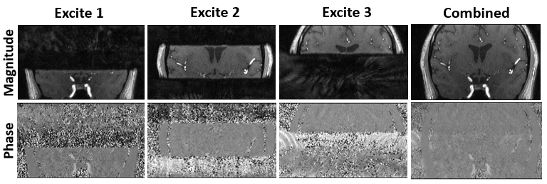
The proposed 4D-flow scans were performed by sequentially exciting thin overlapping slabs. Each slab is acquired using undersampled, distributed-spiral trajectories2. These trajectories are similar to stack of spiral sampling but acquire spirals arms continuously along the phase encode direction with golden angle rotations between adjacent arms. This allows for the high sampling and SNR efficiency, and minimal coherent aliasing. MT preparation is performed intermittently to suppress background signal from the brain matter. After reconstruction, images from individual slabs were combined to yield a single image with gradual weighting from one slab to the next in the overlapping regions to minimize slab boundary artifacts. Images were acquired in a 3T MRI scanner (SIGNA Premier, GE Healthcare, WI, USA) with a 48-channel head coil. Multi-slab scans employed 3 slabs with 25% overlap, TE/TR=2.2ms/9.6ms, Venc=100cm/s, flip angle=15°, 0.75x0.75x1mm resolution, 320x320x96mm FOV, and 96 distributed spiral arms per slice phase encoding. MT preparation was performed every 10 TRs with a 8ms fermi RF pulse with a 950° flip angle and 2400Hz offset frequency. For single-slab scans, 5° flip angle was usedResults
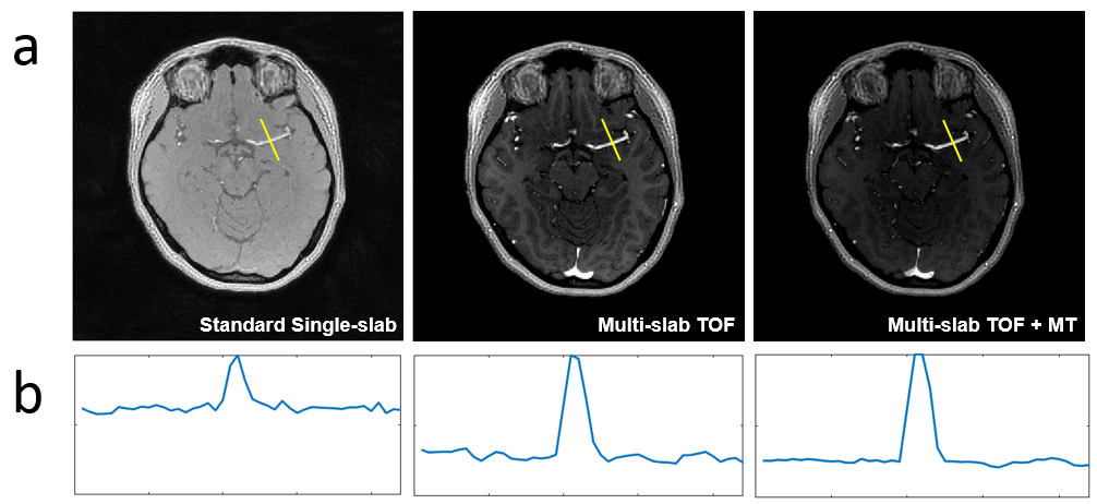
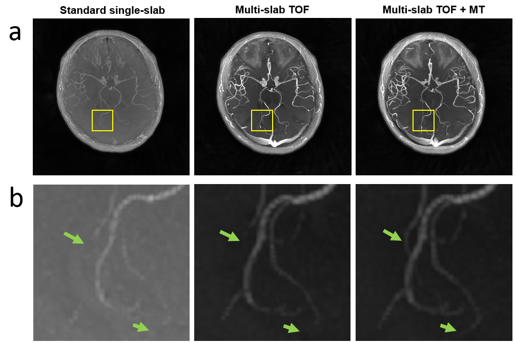
Our approach greatly increased the vessel to background contrast (Fig.2). The multi-slab TOF approach alone produced significant enhancement of vessel signal (Fig.2a,middle) and MT preparation further suppressed brain matter intensity(Fig.2a,right). This resulted in in lower noise profile of the tissue background(Fig.2b,right). Figure 3 shows that our approach was crucial in revealing the vasculature of small vessels in the presence of obscuring background signal from the brain tissue. While most of the background suppression was achieved by the multi-slab TOF approach itself, MT preparation further aids with depiction of some very small vessels (Fig.3b.right) that were otherwise undetectable with TOF approach alone (Fig.3b,middle).Discussion
By combining TOF approach with 4D-flow MRI, we demonstrated a method to improve 4D-flow MRI by utilizing 3D-TOF inflow enhancement in the magnitude images. While 4D-flow MRI is often reliably used to generate MR angiogram and quantify blood flow in large vessels, flow quantification and depiction of smaller vessels are often challenging with standard 4D-flow MRI. Further, in standard 4D-flow MRI, saturation from repeated whole-volume RF excitations lead to gradual signal decay towards the superior part of the brain. As we demonstrated here, the enhanced vessel contrast using the multi-slab approach with MT preparation will aid 4D-flow analysis with more reliable flow quantification and improved vessel segmentation, especially in small vessels.Acknowledgements
No acknowledgement found.References
1. Parker DL, Yuan C, Blatter DD. Angiography by Multiple Thin Slab 3D Acquisition. MRM. 1991. 17:434-451
2. 2. Turley DC, Pipe JG. Distributed spirals: a new class of three-dimensional k-space trajectories. 2013. 70(2):413-9
Figures