2926
Is cerebral blood flow affected by preventable neurovascular risk factors and physical fitness level?1Radiology, University Medical Center Utrecht, Utrecht, Netherlands, 2Danish Research Centre for Magnetic Resonance, Centre for Functional and Diagnostic Imaging and Research, Copenhagen University Hospital Hvidovre, Hvidovre, Denmark, 3Department of Orthopedic Surgery M, Institute of Sports Medicine Copenhagen, Bispebjerg and Frederiksberg University Hospitals, Copenhagen, Denmark, 4Centre for Healthy Aging, Faculty of Health and Medical Sciences, Copenhagen University, Copenhagen, Denmark, 5Department of Public Health, Copenhagen University, Copenhagen, Denmark
Synopsis
The health-related effects of aging (biological age) are highly variable across individuals with a similar chronological age. In this study we investigate the relationship between neurovascular health, physical fitness and mean gray matter cerebral blood flow (CBF) in a cohort of 300 home-dwelling volunteers between 62 and 70 years old. In these population-based elderly participants we found that preventable risk factors for neurovascular disease are associated with cerebral blood flow. A high mean arterial pressure, high cholesterol and low creatinine were associated with a decreased gray matter perfusion, whereas physical fitness was not.
Introduction
Aging is associated with a decline in cognitive ability, physical ability, brain volume, and cerebral blood flow. When reaching the sixth decadethe accumulated effects of environmental, socioeconomic and genetic factors and their interactions are highly variable across individuals.1 In other words, their health and associated predicted mortality differs, which can be referred to as a difference in ‘biological’ age.2 Risk factors for neurovascular disease, including hypertension and diabetes are known to increase biological age.3
In this study we investigate the relationship between characteristics associated with neurovascular health, physical fitness and mean cerebral blood flow (CBF) in cerebral gray matter.
Methods
Participants
The LIve active Successful Aging (LISA) study investigates the relationship between physical fitness, cognition and mental health in volunteers around retirement age.4 People were eligible to participate if they were home dwelling, independent, and between 62 and 70 years of age. Exclusion criteria were participation in a regular exercise regime, presence of severe or dysregulated disease, musculoskeletal diseases impeding training ability, use of systemic glucocorticoids, androgens or antiandrogens and any contraindications for physical exercise or MRI.
Medical examination
Participants were fasting, and asked to refrain from alcohol, and caffeine from 10 pm the evening before the assessments performed by a medical doctor. Height, weight and blood samples were obtained as well as blood pressure in a seated position after at least 5 min rest with an automated oscillometric device. Mean arterial pressure was defined as (2 * Diastolic blood pressure + systolic blood pressure)/3.
Physical tests
Physical fitness was assessed by both muscle strength and cardiorespiratory fitness. Upper limb and lower limb muscle strength were assessed with a grip strength measurement (DHD-1 Digital Hand Dynamometer, Saehan corporation, Seoul, South-Korea) and a leg extensor power test (Leg Extensor Power Rig, Queen’s Medical Centre, Nottingham University, UK), respectively.. Cardiorespiratory fitness was assessed with a 400 meter gait speed test.5 Male and female participants were analyzed separately to account for sex differences in performance on physical tests.
Image protocol and analysis
The MRI protocol of the brain included a 3D-T1w scan (TR/TE=6/2.7 ms, 288×288 matrix, 244 slices, isotropic voxels 0.85mm3) and Arterial Spin Labeling (EPI with SENSE factor 2.5, TR/TE=4610/13 ms, 80×80 matrix, 22 slices, 90 volumes, voxel size 3x3x6 mm3, labeling duration 1800 ms, post labeling delay=2000 ms; M0: TR/TE=9000/13 ms, 22 slices).
Segmentation of gray matter, white matter and cerebrospinal fluid volumes was performed using the 3D-T1w brain scans with FAST tool in FSL (FMBRIB, Oxford, UK). The gray matter map was then registered to ASL space using FSL’s FLIRT tool for linear registration.
ASL quantification was performed with Matlab (2014b). The T1 of blood used in the analysis was corrected individually per subject for hematocrit: T1,blood = 0.52 * hematocrit + 0.38 [6].
Multiple linear regression was performed to identify variables associated with gray matter perfusion.
Results
Three cases were excluded due to a mean gray matter CBF below 20 ml/100g/min. As such, the final analysis included 298 participants (Table 1). There was no difference in mean gray matter CBF between male (45.6 ml/100g/min) and female participants (44.5 ml/100g/min) (P = 0.081).
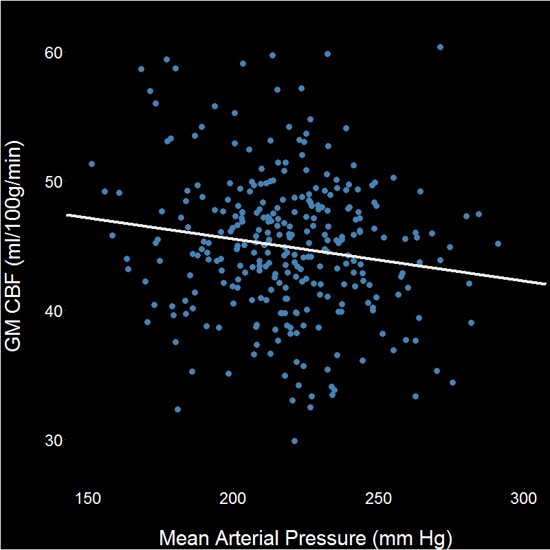
Gray matter CBF was associated with several (neuro)vascular risk factors. High mean arterial pressure (Figure 1), high total cholesterol, and low creatinine were associated with lower cerebral blood flow (Table 2).
Subsequently, performance on the physical tests were added separately to the model to detect any contributions to the explained variance. Performance on any of the physical tests was not associated with perfusion when included as an additional variable in the model for both male and female subjects.
Discussion and conclusion
In population-based elderly participants preventable risk factors for neurovascular disease are associated with cerebral blood flow. A high mean arterial pressure, high cholesterol and low creatinine were associated with a decreased gray matter perfusion.
Leg extensor power and hand grip strength as proxies for lower and upper limb muscle strength and 400m gait speed as a proxy for cardiorespiratory fitness were not associated with mean gray matter perfusion. Future analyses will include regional perfusion measures and cognition.
Acknowledgements
European Research Council under the European Union's Horizon 2020 Programme (H2020) / ERC grant agreement n°637024 (JH).References
1. Mitnitski, A., S.E. Howlett, and K. Rockwood, Heterogeneity of Human Aging and Its Assessment. The Journals of Gerontology: Series A, 2017. 72(7): p. 877-884.
2. Chen, B.H., et al., DNA methylation-based measures of biological age: meta-analysis predicting time to death. Aging (Albany NY), 2016. 8(9): p. 1844-1859.
3. Pyrkov, T.V., et al., Extracting biological age from biomedical data via deep learning: too much of a good thing? Scientific Reports, 2018. 8(1): p. 5210.
4. Eriksen, C.S., et al., Physical activity as intervention for age-related loss of muscle mass and function: protocol for a randomised controlled trial (the LISA study). BMJ Open, 2016. 6(12).
5. Simonsick, E.M., E. Fan, and J.L. Fleg, Estimating Cardiorespiratory Fitness in Well-Functioning Older Adults: Treadmill Validation of the Long Distance Corridor Walk. Journal of the American Geriatrics Society, 2006. 54(1): p. 127-132.
6. Lu, H., et al., Determining the longitudinal relaxation time (T1) of blood at 3.0 Tesla. Magnetic Resonance in Medicine, 2004. 52(3): p. 679-682.
Figures
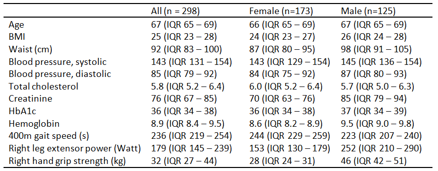
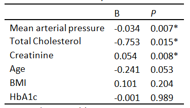
Table 2. Model 1: Gray matter CBF and vascular risk factors.
Dependent
variable = mean gray matter perfusion;
R2
= 9%. F = 4.6. P < 0.0002;