2924
Structural connectivity alterations in CSVD patients with mild cognitive impairment: an atlas-based DTI structural connectome study1Department of Radiology, Tongji Hospital, Tongji medical college, Huazhong University of science and techology, Wuhan, China
Synopsis
Cerebral small vessel disease(CSVD) is the most common cause of vascular cognitive impairment. Early diagnosis and intervention could prevent patients from progressing to dementia rapidly. DTI deterministic tractography and structural network analysis based on graph theory were utilized to explore the difference of structural connectome between CSVD patients and normal individuals. The results demonstrated the disruption of structural connection at global level and reduction of network efficiency at nodal level. Especially, the decrease of nodal efficiency in the right cuneus gyrus was associated with the mild memory loss in CSVD patients. Our results may provide crucial clues to therapeutic interventions.
Introduction
Mild cognitive impairment, characterized by decreased memory, reduced language fluency, and decreased executive ability, etc., is a main cause of hospitalization for CSVD patients. Early diagnosis and intervention prevent patients from progressing into dementia. Recent findings showed the disruption of the cerebral connection and ultimately led to specific symptoms in CSVD patients with cognitive impairment1, However, the specific brain regions associated with mild cognitive impairment has not been clearly elucidated yet. DTI deterministic tractography and structural network analysis based on graph theory2 made it possible to quantitatively evaluate brain network connectivity. Our study aims to investigate the network connection state in CSVD and find out brain regions related to mild cognitive impairment of CSVD patients.Method
Thirty-seven CSVD patients with mild cognitive impairment patients, thirty-four normal control (NC) volunteers were enrolled. White matter pathways were traced among whole brain segmenting into 64 regions derived from JHU-DTI-atlas (https://mricloud.org)to characterize interregional connections based on graph theory. Small-world, global efficiency and local efficiency were computed to characterize the information transfer across a network and locally communicated within neighborhood. At a regional level, betweenness centrality was calculated to identify nodes of a network that play crucial roles in efficient communication, Local node efficiency indicated how well the information can be communicated within the neighbors of a given node when this node was removed.Result
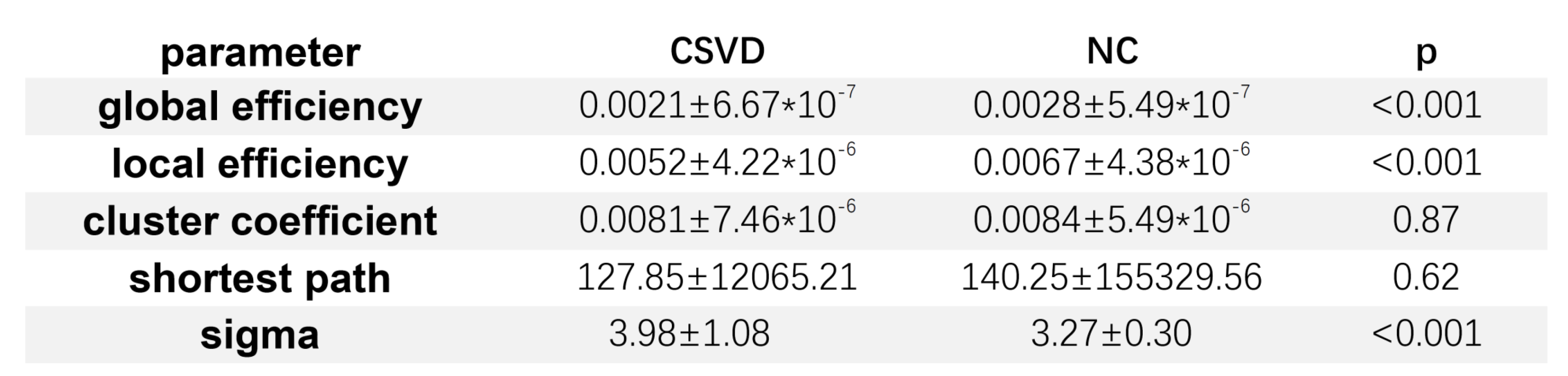
Global level: the brain network in two groups all demonstrated small world properties(γ≥1 and λ ≈ 1 or γ/λ>1)3, suggesting the tenet of most efficient connection has not been destroyed, but the difference of shortest path and cluster coefficient between two groups were not significant. CSVD group displayed significantly lower global efficiency and local efficiency compared with NC group(Figure 1).
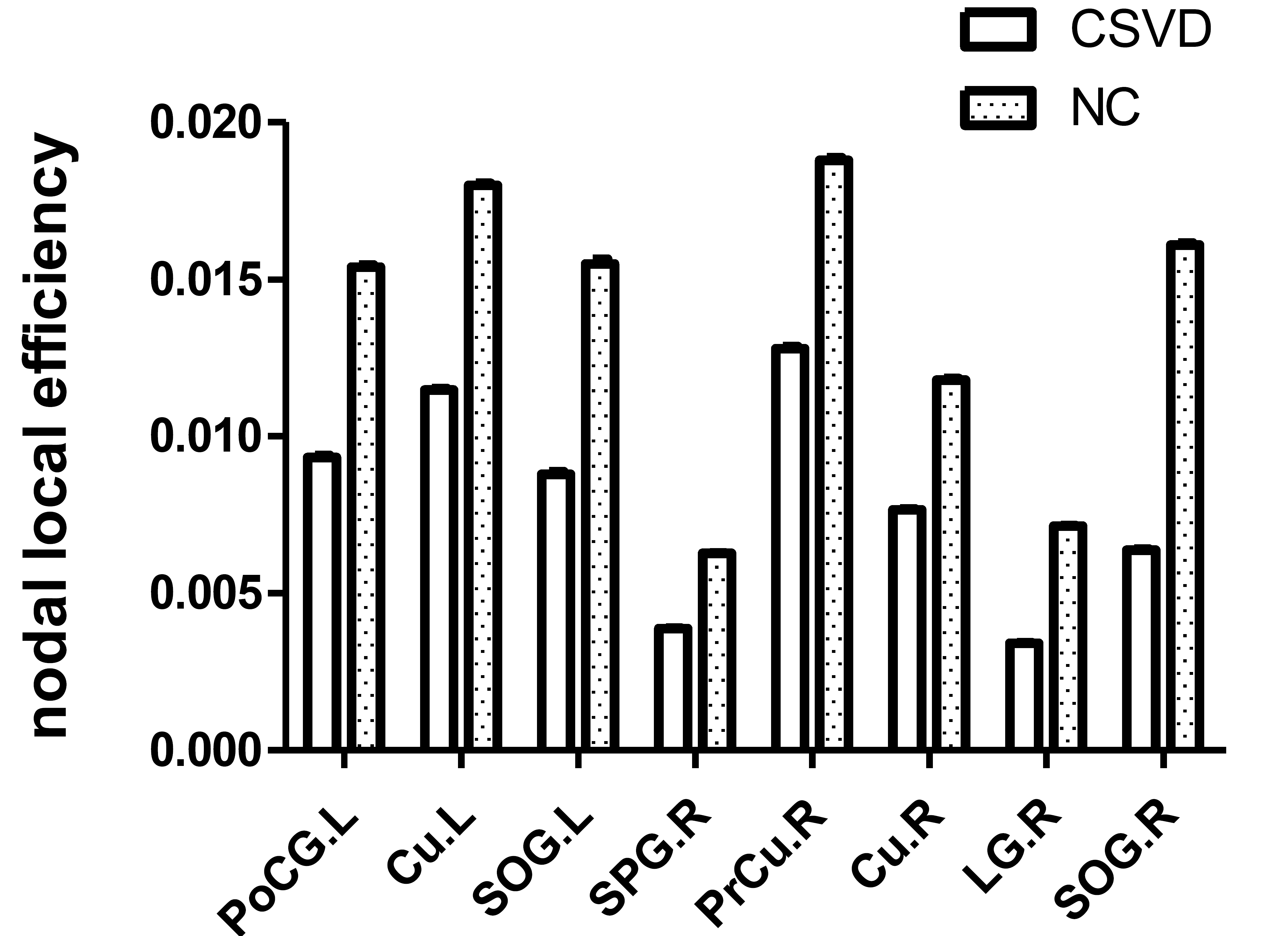
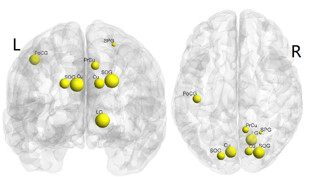
Nodal level: Figure2,3 illustrated the brain regions with significant differences of nodal local efficiency between two groups. Compared to NC group, CSVD group showed lower nodal efficiency in 8 nodes including bilateral cuneus gyrus, superior occipital gyrus, left postcentral gyrus and right pre-cuneus gyrus ,lingual gyrus and superior parietal gyrus (p<0.05, FDR-corrected). No region was found different in betweenness centrality.
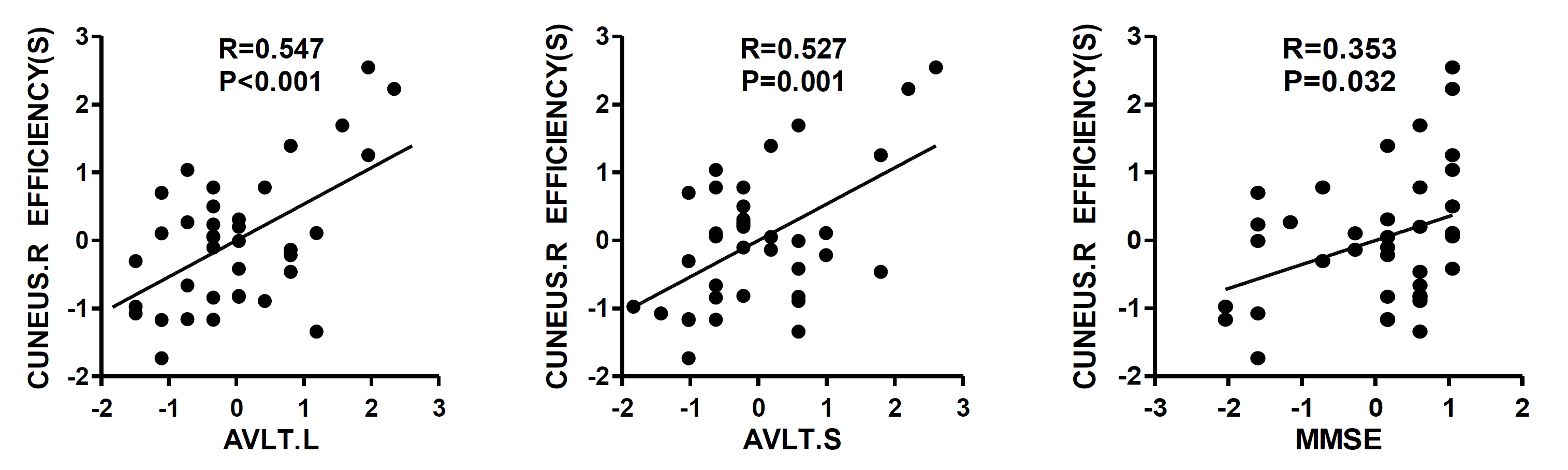
Clinical scales: MMSE, AVLT(S/L), TMT(A/B) and VFT scales were evaluated to quantify overall cognitive function, memory, execution and verbal frequency respectively, only VFT showed no difference between two groups, Moreover, the nodal efficiency of the right cuneus gyrus is positively correlated with AVLT(S/L) and MMSE scores in 37 patients, suggesting decreased nodal local efficiency of the right cuneus gyrus related to an impairment of episodic memory in patients with CSVD.(Figure4)
Discussion
In this study, both groups exhibited small-world characteristics but the difference didn’t differ. XF Xie et al. found patients with depressive symptoms in CSVD also showed small world properties4, as well as in infancy2, which suggested that small world is an inherently property to keep brain in an efficient and tight connection.
CSVD group demonstrated lower efficiency at global level and also in eight brain regions, suggesting that the ability of information integration and separation was weakened in CSVD patients5, in line with the study of Andrew J6. Most of the regions were located around the intra-parietal sulcus and the frontal regions were spared, suggested that the higher-level cognitive occurred after posterior awareness arose7 has not been impaired, corresponding to the very mild cognitive impairment of the CSVD patients enrolled.
Betweenness centrality , defined as the fraction of all shortest paths that pass through the node8,representing the importance of a node’s functional capability in sending signals and passing information in the brain, showed no difference in all regions. However, Seo EH observed that the betweenness centrality was lower in patients with MCI and PD9. The discrepancy may be resulted by the heterogeneity of the patients, or may also suggested that the nodal efficiency a more sensitive index than betweenness centrality assessing the brain structural connectivity as Rubinov M8 reported.
Moreover, episodic memory assessed by AVLT(S/L) was positively correlated with the nodal efficiency changes of the right cuneus gyrus. The decline of nodal efficiency in cuneus gyrus may be caused by the fiber loss connecting to the dorsal frontal cortex10, due to WMH and lacunes which are two important characteristics of CSVD patients, and typically resulted episodic memory loss in those patients11, 12.
Conclusion
Diffusion tensor based on atlas detected network connection by trancing white matter, which has been robustly used in many other diseases. Our study found the reduced network connection in CSVD at both global and node level, as well as an significant association with episodic memory. Our results may provide crucial clues to therapeutic interventions early in CSVD patients.Acknowledgements
Supported by the Key Program National Natural Science Foundation of China (Grant No. 81730049)References
[1] Benarroch E E: The midline and intralaminar thalamic nuclei, Neurology, 2008, 71(12): 944-949. [2] Ratnarajah N, Rifkin-Graboi A, Fortier M V, et al: Structural connectivity asymmetry in the neonatal brain, NeuroImage, 2013, 75: 187-194. [3] Watts D J, Strogatz S H: Collective dynamics of ‘small-world’ networks, Nature, 1998, 393: 440. [4] Xie X, Shi Y, Zhang J: Structural network connectivity impairment and depressive symptoms in cerebral small vessel disease, Journal of Affective Disorders, 2017, 220: 8-14. [5] Makovac E, Mancini M, Fagioli S, et al: Network abnormalities in generalized anxiety pervade beyond the amygdala-pre-frontal cortex circuit: Insights from graph theory, Psychiatry Research: Neuroimaging, 2018, 281: 107-116. [6] Lawrence A J, Chung A W, Morris R G, et al: Structural network efficiency is associated with cognitive impairment in small-vessel disease, Neurology, 2014, 83(4): 304-311. [7] Koivisto M, Ruohola M, Vahtera A, et al: The effects of working memory load on visual awareness and its electrophysiological correlates, Neuropsychologia, 2018, 120: 86-96. [8] Rubinov M, Sporns O: Complex network measures of brain connectivity: Uses and interpretations, NeuroImage, 2010, 52(3): 1059-1069. [9] Seo E H, Lee D Y, Lee J, et al: Whole-brain Functional Networks in Cognitively Normal, Mild Cognitive Impairment, and Alzheimer’s Disease, PLOS ONE, 2013, 8(1): e53922. [10] Goldman-Rakic P S: Topography of cognition: Parallel Distributed Networks in Primate Association Cortex, Annual Reviews Inc, 1988, 11: 137-156. [11] Cabeza R, Mangels J, Nyberg L, et al: Brain Regions Differentially Involved in Remembering What and When: a PET Study, Neuron, 1997, 19(4): 863-870. [12] Owen A M, Milner B, Petrides M, et al: A Specific Role for the Right Parahippocampal Gyrus in the Retrieval of Object-Location: A Positron Emission Tomography Study, Journal of Cognitive Neuroscience, 1996, 8(6): 588-602.Figures