2921
Towards quantitative characterization of brain tumors using synthetic MRI: a preliminary study with pathological confirmationHui-ming LIU1, Tie-bao MENG1, Hao-qiang HE1, Liangru Ke1, Long Qian2, Bing Wu2, Guo-ping Yin3, and Chuan-miao XIE1
1Department of Medical Imaging, Sun Yat-sen University Cancer Center, Guangzhou, China, 2GE Healthcare, MR Research China, Beijing, China, 3GE Healthcare, MR Application China, Beijing, China
Synopsis
MRI is widely involved for the diagnosis of brain tumors, and offers qualitative assessment of the tumor with regard to the surrounding brain tissues. However, not only visual inspections may introduce subjective bias, there may be cases that pose challenges for diagnosis even with the use of contrast agent. In this work, a novel relaxation quantification approach named synthetic MRI was applied to 40 patients with glioma or meningioma. Our data showed that both T1 and T2 values of glioma were significant higher than that of meningioma. indicating that synthetic MR may be used for classifying different types of brain tumors.
Introduction
MRI is widely involved for the diagnosis and prognosis of brain tumors, and T1, T2 and PD weighted images along with diffusion weighted images are routinely used sequences. Theses sequences offer qualitative assessment of the tumor with regard to the surrounding brain tissues for clinical decisions. Not only visual inspections may introduce subjective bias, there may be cases that pose challenges for diagnosis even with the use of contrast agent, such as metastases and high-grade glioma. It has been reported that different types of brain tumors feature distinctive T1 values 1, it is envisioned if an atlas that associates the pathological properties of brain tumors with magnetic relaxation times may be built, diagnosis of brain tumors may be more objective and accurate, and it may also eliminate the needs for contrast agents. Synthetic MRI offers a good solution for such needs, where multiple contrasts and relaxation time maps are simultaneously obtained. This work reports part of the ongoing efforts in quantitative characterization of brain tumors using multiple relaxation times.Methods
Local ethical approval and consent forms were obtained for this project. From January 6th to August 28th, 2018, a total of 89 patients suspected with brain tumors were enrolled in this prospective study. The inclusion criteria were receiving surgical operations and pathological confirmations were either glioma or meningioma; exclusion criteria were unsuccessful scans due to incorporative patients. All the patients underwent MR exams on a 3.0T whole body scanner (Signa Pioneer, GE, WI) equipped with an 8-chanel head coil, the MR scans included both conventional sequences as well as synthetic MR (magnetic resonance image compilation, MAGiC). The scan parameters for synthetic MR were: TR = 4000 ms, TE1=21 and TE2=95 ms, slice thickness/gap = 5/1 mm, FOV = 28 cm, data matrix = 320*256, echo length = 16, bandwidth = 41.67 Hz. Region of interests (ROI) were placed on the lesions based on its contrast enhanced images by an experienced radiologist, then the ROIs were copied onto T1/T2 relaxation maps and mean relaxation values were recorded. The results were then analyzed by independent sample t-test.Results
Figure 1 shows the representative images of glioma and meningioma from synthetic MR, including both contrast weighted images and T1/T2 relaxation maps. The T1 and T2 values in patients with glioma and meningioma are shown in Table 1. Both T1 and T2 values of glioma were significant higher than that of meningioma (P<0.05). The pathological results of glioma and meningioma are shown in Figure 2, respectively. ROC analysis of T1 values showed that the area under curve (AUC) was 0.875 and the cut-off value was 1435 ms with specificity and sensitivity equal to 0.722 and 1, respectively. With regard to T2 map, the AUC was 0.903 and the cut-off value was 113 ms, where the specificity and sensitivity equal to 0.667 and 1, respectively (Figure 3).Discussion
T1 and T2 relaxations may reflect the tissue composition and hence have the potential to be quantitative biomarkers for different pathological properties. Past work has reported relaxation association with the brain tumor types 1, however the approach only focused on T1 relaxation time and was not suited for clinical use due to scan time and may be biased by B1 inhomogeneity. With the availability of synthetic MR, multiple relaxation times may be obtained with clinical acceptable time and B1 correction. In this preliminary work, it was seen that glioma and meningioma feature distinctive relaxation times. Continuous accumulation of other types of brain tumors and pathological confirmations may allow quantitative differential diagnosis in the future.Conclusion
Synthetic MR may be used for building quantitative relaxation based characterization for different types of brain tumors.Acknowledgements
No acknowledgement found.References
1. Araki, T., et al. "Magnetic resonance imaging of brain tumors: measurement of T1. Work in progress." Radiology 150.1 (1984): 95-98.Figures
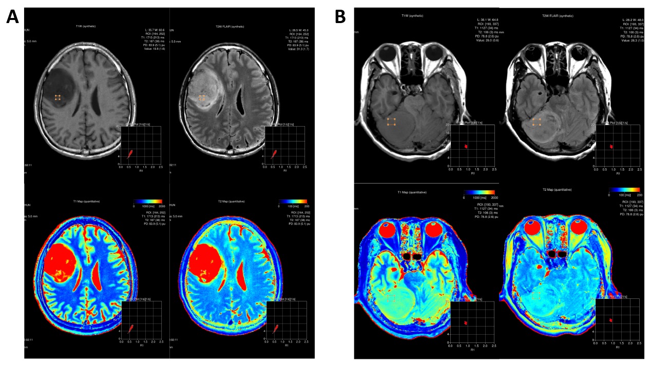
Figure 1. A, male, represents a case with glioma, male, WHO III. The
T1 and T2 value of the lesion were 1713 and 187 ms, respectively. B, female, represents
a subject with meningioma, WHO I. The T1 and T2 value of the lesion were 1127
and 106 ms, respectively.
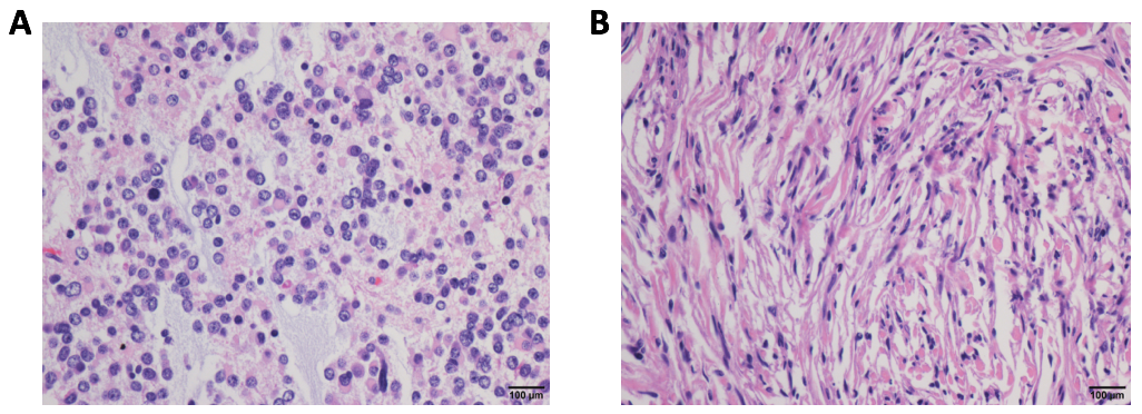
Figure 1. The
pathological results of glioma (A) and meningioma (B) corresponding to the
cases of Figure 1.
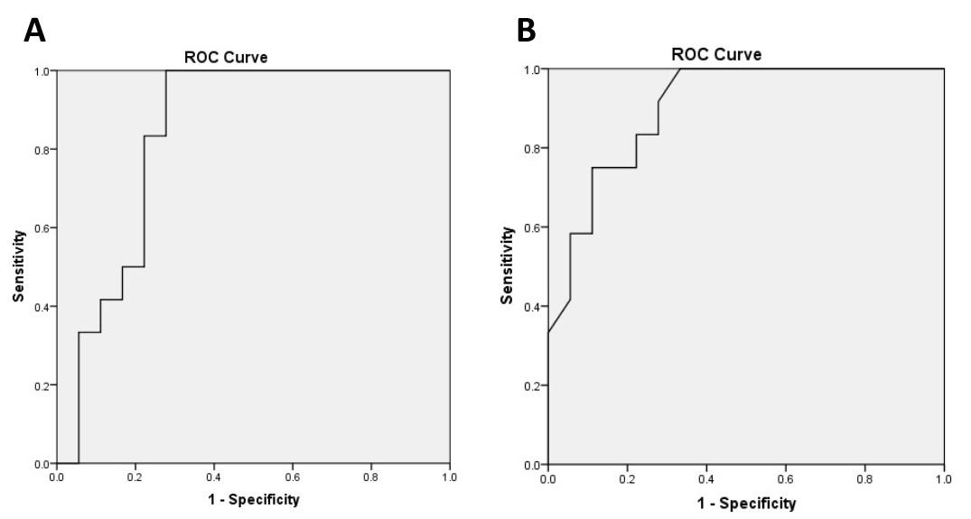
Figure 3. A represents the ROC curve of T1 value, the area under curve
(AUC) was 0.875 and the cut-off value was 1435 ms with specificity and
sensitivity equal to 0.722 and 1, respectively.
B represents the ROC curve of T2 value, the
AUC was 0.903 and the cut-off value was 113 ms, where the specificity
and sensitivity equal to 0.667 and 1, respectively.