2918
The Value of Intravoxel Incoherent Motion Imaging in Predicting the Survival of Patients with Astrocytoma1Shanxi Cardiovascular Hospital, Taiyuan, China, 2the First Hospital of Shanxi Medical University, Taiyuan, China, 3GE Healthcare, MR Research, Beijing, China
Synopsis
In this study, two independent sample t-tests was used to evaluate the predictive value of IVIM parameters for the two-year survival rate of 60 patients with astrocytoma, and the correlation between IVIM parameters and survival days was analyzed by Pearson correlation. The results showed that the ADC, D*, f value of IVIM parameters had great potential in predicting the two-year survival rate of astrocytoma patients, which was related to survival days.
Objective
MR multi-b value diffusion weighted imaging (DWI) combined with bi-exponential models (intravoxel incoherent motion imaging, IVIM) can be used to assess the biological behavior of tumor in both diffusion and microcirculation perfusion1, which was originally applied in the grading of gliomas2. However, Application of IVIM in the prognosis of gliomas has rarely been reported. Astrocytome is one of the representatives of glioma. The aim of this study is to explore the value of IVIM in predicting the survival rate of patients with cerebral astrocytoma.Methods
Sixty patients (males: females = 34: 26, mean age 62.23±16.32 years) with cerebral astrocytomas confirmed by pathology underwent MR scans of multi-b value DWI on a 3.0 Tesla GE Signa HDxt MRI scanner (GE, Milwaukee, WI, USA) with 8-channel head and neck combined coil. Patients were divided into a death group and a survival group after a two-year follow-up. Regions of interest were chose in the tumor parenchyma. Values of quantitative parameters in multi-b value DWI, including ADC, D, D* and f, were calculated. Two-sample t-tests were employed to compare the parameters between the two patient groups. The diagnostic performance of the parameters was assessed by the Receiver Operating Characteristic (ROC) curve analysis. Correlations between the quantitative parameters and the survival days were analyzed through Pearson correlation analysis.Results and Discussion
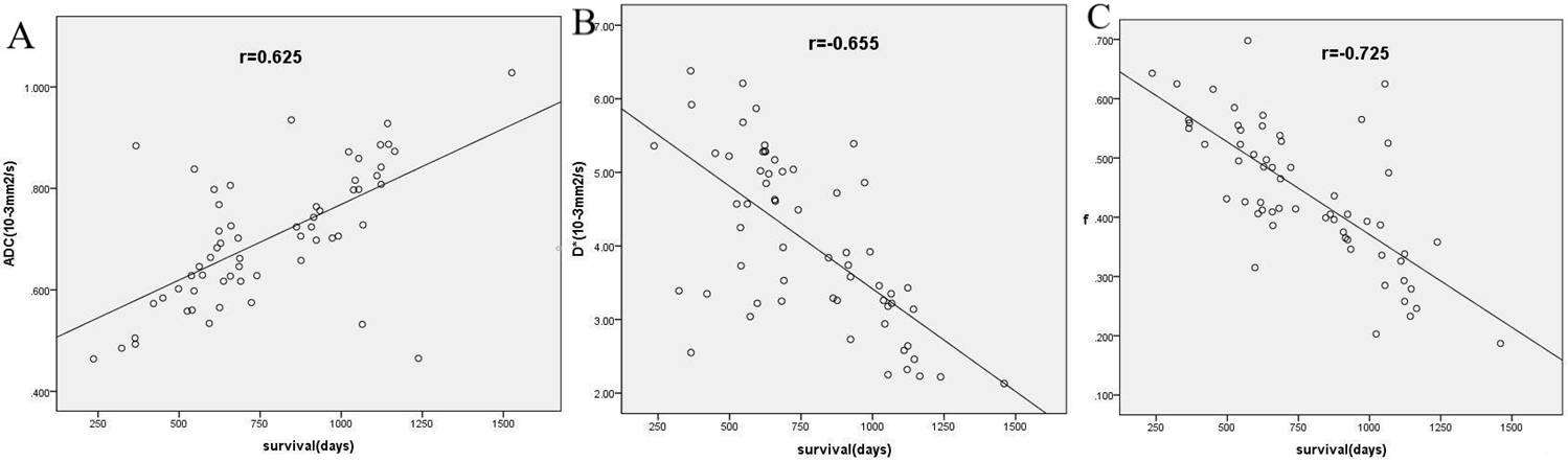
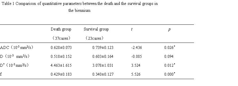
Except the D value (p = 0.094), all other the parameters demonstrated a great statistical difference between the death and the survival group in Table 1. According the literatures3,4, the ADC value can reflect the tumor cell density, and the D* value and f value represent micro-vessel formation. The lower the ADC value is, the greater the tumor cell density is. And the greater the D* and f values, the more micro-vessel formation. The proliferation of tumor cell density and micro-vessel may predict the non-ideal prognosis of patients. The area under the ROC curve of the ADC, D*, f were 0.811, 0.858, 0.892, respectively, which indicated a higher accuracy of f value in predicting the two-year survival rate when compared to ADC and D* values. D* value was higher than ADC, because many studies5, 6 have shown that the attenuation of in vivo tissue signal depends not only on the diffusion of water molecules, but also on the blood flow of microcirculation in capillaries. ADC values cannot be used to distinguish diffusion and perfusion, so it cannot accurately reflect the complex structure of the biological tissue and the movement of its molecules. As for the D* and f values, the multi-component perfusion have more influence on D* value than on f value. The ADC in Fig 1-A (r = 0.625, p = 0.023), D* in Fig1-B (r = -0.655, p = 0.012) and f in Fig 1-C (r = -0.725, p = 0.000) were closely correlated with the survival days.Conclusion
The IVIM technique demonstrated great potential in predicting the two-year survival rate for patients with astrocytoma, which may benefit future individualized treatment.Acknowledgements
We are grateful for the National Natural Science Foundation of China (grant No. 21072120), the Shanxi Patent Promotion Implementation Project (20161001), and Key Research and Development (R&D) Projects of Shanxi Province(201703D321008).References
1 Federau C, Cerny M, Roux M, et al. IVIM perfusion fraction is prognostic for survival in brain glioma.Clin Neuroradiol.2017;27(4):485-492.
2 Osamu T, Akio H, Koji Y, et al. Differentiation of high-grade and low-grade diffuse gliomas by intravoxel incoherent motion MR imaging.Neuro Oncol.2016;18(1):132-141.
3 Chen SD, Hou PF, Lou L, et al. The correlation between MR diffusion-weighted imaging and pathological grades on glioma.Eur Rev Med Pharmacol Sci.2014;18(3):1904-1909.
4 Deike K, Wiestler B, Graf M, et al. Prognostic value of combined visualization of MR diffusion and perfusion maps in glioblastoma.J Neurooncol.2016;126(3):463-472.
5 Bisdas S,Koh TS,Roder C,et al. Intravoxel incoherent motion diffusion-weighted MR imaging of gliomas: feasibility of the method and initial results. Neuroradiology.2013;55(10):1189–1196.
6 Wetscherek A,Stieltjes B,Laun FB,et al. Flow-compensated intravoxel incherent motion diffusion imaging.Magn Reson Med.2015;74(2):410-419.
Figures