2912
Noninvasive IDH1 genotype prediction in grade Ⅱ/Ⅲ gliomas based on conventional MR images: a transfer learning strategy1Department of Radiology, Department of Radiology & Functional and Molecular Imaging Key Lab of Shaanxi Province, Xi’an, China
Synopsis
Purpose: To evaluate the performance of transfer learning with CNNs in predicting IDH1 genotype. Method and Materials: AlexNet, GoogLeNet, ResNet and VGGNet were pre-trained on the large scale natural image database (ImageNet) and fine-tuned with T1CE and FLAIR images. The outputs of training set were utilized to train LR and SVM models. Besides, fused images combining FLAIR and T1CE were used to fine-tune pre-trained ImageNet models. Results: Performances were improved by fine-tuning the four architectures with fused images. Conclusion: Transfer learning with various CNNs (especially VGGNet) is powerful in predicting IDH1 genotype in grade Ⅱ/Ⅲ gliomas.
INTRODUCTION
Isocitrate dehydrogenase 1 (IDH1) genotype is highly related with the diagnosis, treatment and prognosis of gliomas, especially in World Health Organization (WHO) grade Ⅱ/Ⅲ gliomas1-3. Pre-treatment identification of IDH1 genotype is extremely helpful in deciding the most appropriate therapeutic strategy. Currently, IDH1 genotype cannot be determined without obtaining tissue samples via invasive surgical procedures. Nevertheless, magnetic resonance (MR) imaging phenotypes have been demonstrated promising noninvasive surrogates for tumor genotypes in recent studies4. And transfer learning has shown its potential in medical imaging analysis5. This study was to evaluate the performance of transfer learning with convolutional neural networks (CNNs) in predicting the IDH1 genotype in grade Ⅱ/Ⅲ gliomas.METHODS
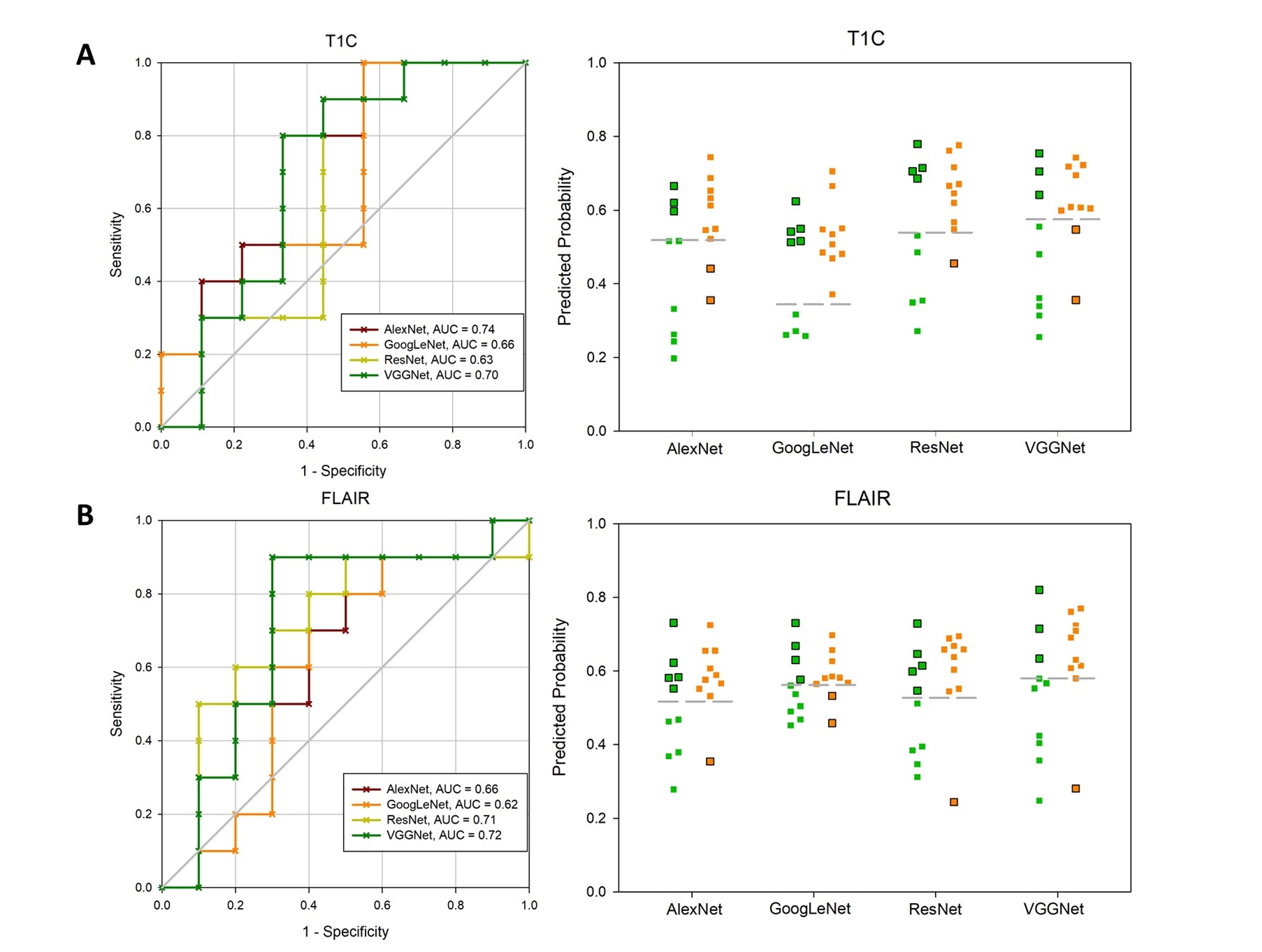
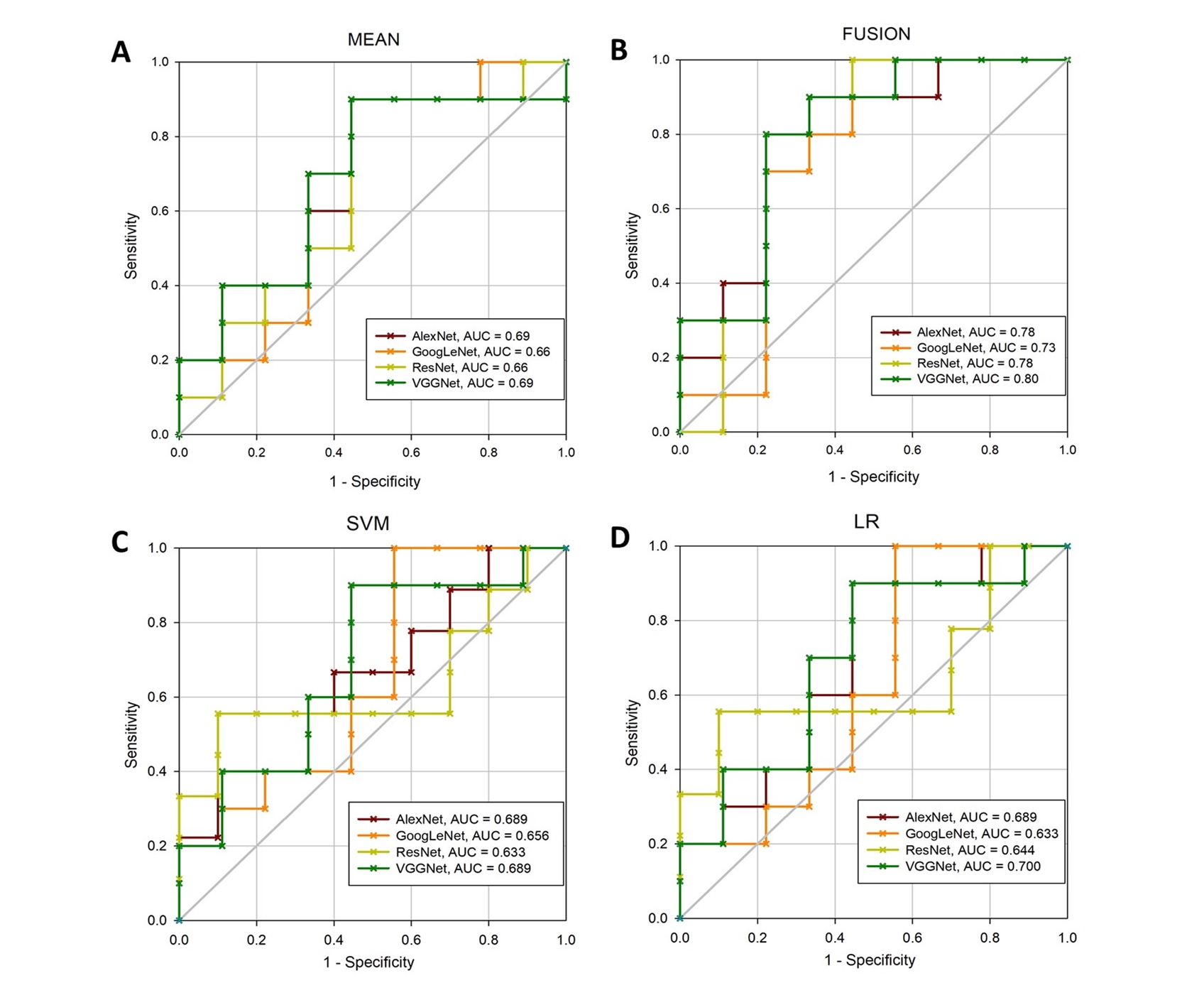
110 pathological confirmed grade Ⅱ/Ⅲ glioma patients were retrospectively included in the current study. For each patient, preoperative conventional magnetic resonance images (MRIs), i.e. fluid attenuation inversion recovery (FLAIR) and contrast-enhanced T1-weighted images (T1CE), were acquired. Four CNNs, including AlexNet, GoogLeNet, ResNet and VGGNet, were pre-trained on the large scale natural image database (ImageNet) and fine-tuned with T1CE and FLAIR images. The outputs of training set were utilized to train logistic regression (LR) and support vetor machine (SVM) models. Besides, fused images combining FLAIR and T1CE were used to fine-tune pre-trained ImageNet models. Predictive performance was assessed by receiver operating characteristic (ROC) curve analysis.RESULTS
Within AlexNet, GoogLeNet, ResNet and VGGNet model, area under the ROC curve (AUC) of IDH1 prediction achieved 0.660, 0.620, 0.710, 0.720 for FLAIR and 0.744, 0.656, 0.633, 0.700 for T1CE images, respectively. Improved performances were obtained by fine-tuning the four architectures with fused images and the AUC reached to 0.778, 0.733, 0.778 and 0.800, respectively.CONCLUSION
Transfer learning with various CNNs (especially VGGNet) is powerful in predicting IDH1 genotype in grade Ⅱ/Ⅲ gliomas. Thus, our procedure is promising to facilitate presurgical molecular pathological diagnosis.Acknowledgements
We would like to thank Dr. Xiao-Cheng Wei from GE healthcare for providing technical support regarding the appropriate amide proton transfer weighted imaging and data analysis.References
1. Schumacher T, Bunse L, Wick W, et al: Mutant IDH1: An immunotherapeutic target in tumors. Oncoimmunology 3:e974392, 2014.8.
2.Rohle D, Popovici-Muller J, Palaskas N, et al: An inhibitor of mutant IDH1 delays growth and promotes differentiation of glioma cells. Science 340:626–630, 2013.9.
3.Schumacher T, Bunse L, Pusch S, et al: A vaccine targeting mutant IDH1 induces antitumour immunity. Nature 512:324-327, 2014.
4. Smits M, van den Bent MJ: Imaging Correlates of Adult Glioma Genotypes. Radiology 284:316-331, 2017.
5.Shin HC, Roth HR, Gao M, et al: Deep Convolutional Neural Networks for Computer-Aided Detection: CNN Architectures, Dataset Characteristics and Transfer Learning. IEEE Trans Med Imaging 35:1285-1298, 2016.
Figures