2908
Diagnostic accuracy of serial 99mTc MDM (bis-Methionine-DTPA) SPECT imaging in differentiation of glioma recurrence from radiation Necrosis: A comparative study with Dynamic Susceptibility Contrast-Enhanced (DSCE)-MRI and Magnetic Resonance Spectroscopy (MRS)Nisha Rani1, Prof Baljinder Singh1, Dr Naredra Kumar2, Dr Paramjeet Singh3, Dr Anil Kumar Mishra4, and Dr Puja Hazari4
1Nuclear medicine, Postgraduate Institute of Medical Education and Research, Chandigarh, India, 2Radiotherapy, Postgraduate Institute of Medical Education and Research, Chandigarh, India, 3Radiodiagnosis and Imaging, Postgraduate Institute of Medical Education and Research, Chandigarh, India, 4Division of Cyclotron and Radiopharmaceutical Sciences, INMAS DRDO, Delhi, India
Synopsis
The use of complimentary imaging modalities in the glioma management of individual patients can provide additional information for further treatment strategy. In view of the limitations of anatomical imaging, cumbersome radio labeling procedures of PET tracers with amino-acids, in the present study, we have performed multimodality imaging using 99mTc-methionine SPECT (as cost effective substitute for expensive amino acid PET imaging), perfusion MRI and MRS for the the differentiation of radiation necrosis from recurrent/residual glioma by the means of serial imaging.
Abstract
Introduction: The current approach of the use of combinational radiological techniques and imaging protocols those are designed for making head-to-head comparison, is gradually becoming the ‘standard of care’ during the post –surgical/treatment follow-up period in glioma patients. Moreover, in the follow-up studies, the use of a complementary imaging method (eg, PET or MRS/Perfusion MRI) in the management of individual patients could provide an important additional information on glioma activity 1, 2, 3. In this study, 99mTc MDM (bis-methionine-DTPA) SPECT 4, 5 was used for the detection of recurrent/residual glioma from radiation necrosis and the results were compared with DSCE-MRI, MRS and clinical findings. Methods: This prospective study enrolled 72 cases initially presented with histologically proven glioma and underwent surgical intervention followed by radiotherapy (+/- chemotherapy). Amongst these 72 patients, fifty patients (50/72) were studied in the initial diagnostic phase after resection of the tumor and remaining 22 patients had been treated previously and studied in the recurrent disease phase. Fifty (50/92) patients underwent sequential follow-up for response assessment. These fifty patients underwent both 99mTc-MDM-SPECT and MR imaging procedures at two time points, that is, at 1 or 2 weeks prior radiotherapy/chemotherapy and at 6 months after radiotherapy/concurrent or adjuvant chemotherapy treatment, respectively. Out of 50 patients, twenty-six (26/50) patients underwent an additional follow-up imaging at post-therapy follow-up period at 12 months. Among these 26 patients, eight (8/26) patients had a fourth 99mTc-MDM-SPECT and MR imaging study at the median follow-up period of 22.5 months (range, 18-24 months). The target (lesion) to non-target (T/NT) ratio was evaluated for MDM-SPECT and SPECT/MR fusion was done by appropriate software (Multimodality Oasis Server version 1.9.4.3, Segami Corp.). Relative cerebral blood volume (nCBVmax) maps were calculated using perfusion analysis software (Nordic NeuroLab, Bergen, Norway) and signal intensities of choline (Cho), total creatine (Cr), N-acetyl aspartate (NAA), and lipids-lactate (LL) were analysed to calculate Cho/Cr, Cho/NAA, Cr/NAA, Cr/Cho and Cho/LL ratios. Final diagnosis was based on the follow-up imaging findings (both visual and quantitative) as well as the consensus of multidisciplinary neuro-oncology team based on clinical course. Results: After follow-up in the present study, twenty- five (25/72) patients showed necrosis and remaining 47 patients showed recurrent or residual disease.. Table no.1 illustrating mean ± SD values for recurrent/residual and radiation necrosis for all seven quantitative ratios with their statistical significance. The quantitative ratios (T/NT, nCBVmax , Cho/NAA, Cho/Cr, Cr/NAA, Cr/Cho and Cho/LL) achieved an acceptable balance between sensitivity and specificity for distinguishing recurrent tumor to radiation necrosis and each ratio defined a cut off for diagnosing recurrent tumor. The ratios are as follows: T/NT> 1.90 (sensitivity 97.9 % and specificity 92%, AUC 0.971 ±.02), nCBVmax > 3.32 (sensitivity 84.6%, specificity 93%, 0.932 ±.03), Cho/NAA >1.57 (sensitivity 81%, specificity 73%, AUC 0.714 ±.10 ), Cho/Cr>1.64 (sensitivity 85.3%, specificity 73.7%, AUC 0.758 ±.09), Cr/NAA>1.06 (sensitivity 57.1%, specificity 63.6%, AUC 0.569 ±.10), Cr/Cho ≤0.60 (sensitivity 72.3%, specificity 81%, AUC 0.771 ±.09) and Cho/LL>0.90 (sensitivity 71.4%, specificity 50%). The T/NT ratio showed a positive correlation with CBV, Cho/NAA, Cho/Cr, and Cho/LL (r = 0.775, P <0.00001; r = 0.467, P = 0.007; r = 0.368, P = 0.03 and r = 0.443, P = 0.03), respectively. The metabolite ratios of Cr/NAA showed a trend for correlation with the T/NT ratio; however they did not reach statistical significance (r = 0.185, P = 0.31). The Cr/Cho ratio showed negative correlation with T/NT ratio (r = -0.482, P = 0.005). The T/NT ratio showed strongest linear correlation with CBV followed by Cho/NAA. Discussion: Our results of 99m-Tc MDM SPECT and advance MR quantifications are in consonance with the previous studies 6, 7, 8. Furthermore, in the follow-up of 50 patients with repeat imaging, we observed that any change (increase or decrease) in T/NT ratio was significantly associated with the similar pattern in CBV, MR Spectroscopy metabolite ratios. Conclusion: This study indicates that 99mTc-MDM SPECT/CT and DSCE MRI demonstrated more accurate compared to MRS for the detection of tumor recurrence in glioma patients. Furthermore, this study revealed that co-registered MDM-SPECT and MRI facilitates for identification of regions with recurrence. Advanced imaging 99mTc-MDM SPECT/CT with functional MRI may be a powerful tool for accurate planning further glioma patient management.Acknowledgements
The author (Nisha Rani) is thankful to INMAS, DRDO, Delhi for providing funding to this project as an extramural grant. The author is also thankful to Nordic Neuro Lab, Norway and Segami Corp, Houston, USA for providing us software on trial basis.References
1. Deng SM, Zhang B, Wu YW, Zhang W, Chen YY. Detection of glioma recurrence by 11C-methionine positron emission tomography and dynamic susceptibility contrast-enhanced magnetic resonance imaging: a meta-analysis. Nucl Med Commun. 2013;34(8):758-66. 2. Jenkinson MD, Du Plessis DG, Walker C, Smith TS. Advanced MRI in the management of adult gliomas. Br J Neurosurg. 2007;21(6):550-61. 3. Chen W. Clinical applications of PET in brain tumors. J Nucl Med. 2007;48(9):1468-81. 4. Singh B, Kumar N, Sharma S, Watts A, Hazari PP, Rani N, et al. 99mTc-MDM Brain SPECT for the Detection of Recurrent/Remnant Glioma—Comparison With ceMRI and 18F-FLT PET Imaging: Initial Results. Clin Nucl Med. 2015;40(10):e475-e479. 5. Rani N, Singh B, Kumar N, Singh P, Hazari PP, Singh H, et al. Differentiation of Recurrent/Residual Glioma From Radiation Necrosis Using Semi Quantitative 99mTc MDM (Bis-Methionine-DTPA) Brain SPECT/CT and Dynamic Susceptibility Contrast-Enhanced MR Perfusion: A Comparative Study. Clinical nuclear medicine. 2018 Mar 1;43(3):e74-81. 6. Sadeghi N, Salmon I, Tang BN, et al. Correlation between dynamic susceptibility contrast perfusion MRI and methionine metabolism in brain gliomas: preliminary results. J Magn Reson Imaging. 2006;24:989–994. 7. Sadeghi N, Salmon I, Decaestecker C, et al. Stereotactic comparison among cerebral blood volume, methionine uptake, and histopathology in brain glioma. AJNR Am J Neuroradiol. 2007;28:455–461. 8. Kracht LW, Friese M, Herholz K, et al. Methyl-[11C]-l-methionine uptake as measured by positron emission tomography correlates to microvessel density in patients with glioma. Eur J Nucl Med Mol Imaging. 2003;30:868–873Figures
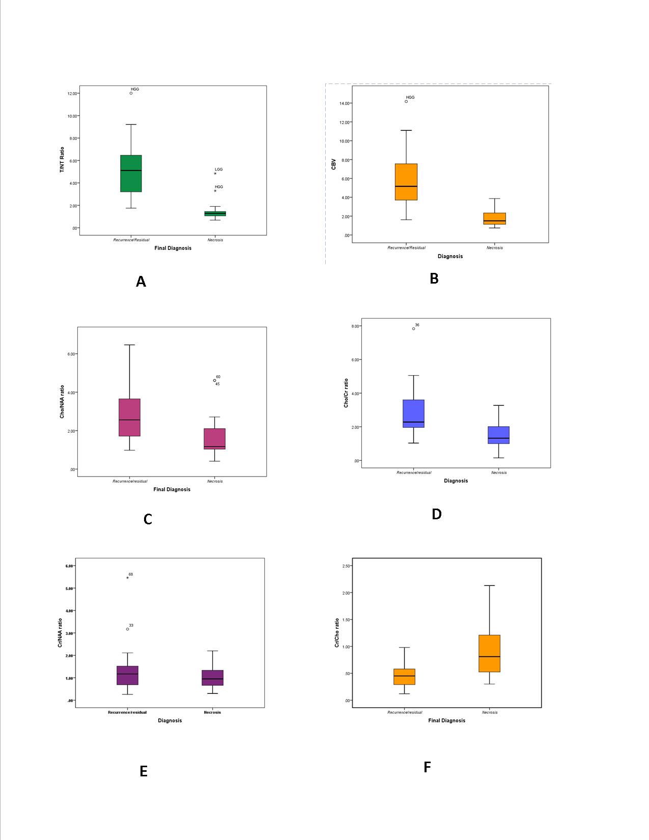
fig1. box and whisker diagrams demonstrating mean ±SD of T/NT ratios MR–derived quantitative values in tumor recurrence versus tumor necrosis.
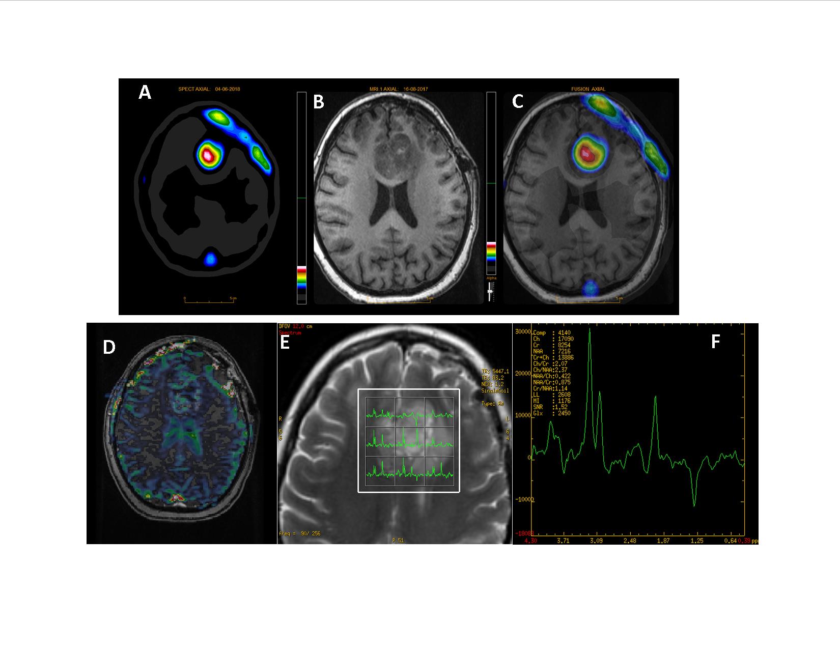
A 37-year-old woman with GBM (G-IV)
at 12 months after surgery/radiotherapy. 99mTc MDM SPECT (transaxial image)
demonstrating foci of increased tracer concentration in the corpus callosum
areas (A). MR image (transaxial,) indicating heterogeneous hyperintense
enhancement in the corresponding area (B). Fused 99mTc MDM SPECT and MR transaxial slices (C). DSCE-MR imaging presenting perfusion maps (D) in the tumor and multivoxel MR Spectra
with increased cho peak demonstrating (E,
F)tumor recurrence.