2907
Restricted ketogenic diet and fasting in combination with re-irradiation in glioblastoma impact intracellular pH and intracerebral metabolism1Institute of Neuroradiology, University Hospital, Goethe-University Frankfurt, Frankfurt, Germany, 2Dr. Senckenberg Institute of Neurooncology, University Hospital, Goethe-University Frankfurt, Frankfurt, Germany, 3Institute of Neurooncology, University Hospital Tübingen, Tübingen, Germany
Synopsis
Inhibition of glycolysis by decreasing blood glucoses levels and increasing ketone bodies (KB) could force tumor cells to shift their metabolism towards potentially impaired mitochondria. The aim of this study was to explore the combination of six days of calorie restricted ketogenic diet (crKD) and three days of fasting (as a possible radiosensitizer) with a re-irradiation therapy in patients with recurrent glioblastoma. Intracerebral concentrations of KB as well as pHi and ATP were non-invasively monitored using MR-Spectroscopy. We were able to show evidence of intratumoral acetone using 1H-MRS in some patients with malignant glioma at day 6 of crKD/fasting. Changes in pHi and ATP during crKD/fasting will remain subject to preclinical studies.
Introduction
Despite multimodal treatment options, glioblastoma continues to carry a poor prognosis. It is therefore of high interest to find combination therapies. While normal neurons and glial cells can metabolize either ketone bodies (KB) or glucose, malignant brain tumor cells might lack this metabolic flexibility due to impaired mitochondrial function (1–3). Inhibition of glycolysis by decreasing blood glucoses levels and increasing KB, could force tumor cells to shift their metabolism towards the impaired mitochondria. Tumors cells maintain a slightly alkaline intracellular pH (pHi) compared to normal cells through changes in the expression of cellular membrane ion transport channels and the CO2/HCO3-buffering system (4). The aim of this study was to explore the combination of six days of calorie restricted ketogenic diet (crKD) and three days of fasting (as a possible radiosensitizer) with a re-irradiation therapy in patients with recurrent glioblastoma. Intracerebral concentrations of KB as well as pHi and ATP were non-invasively monitored using MR-Spectroscopy.Methods
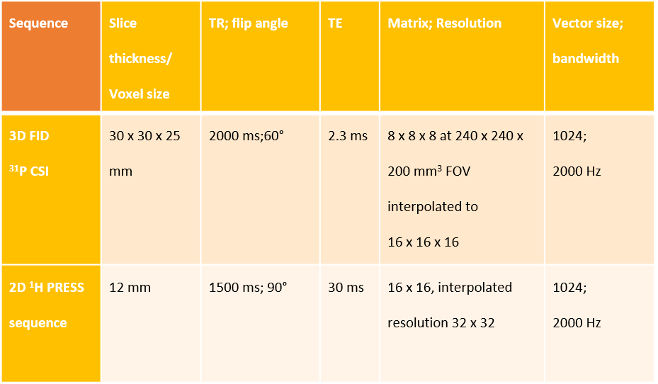
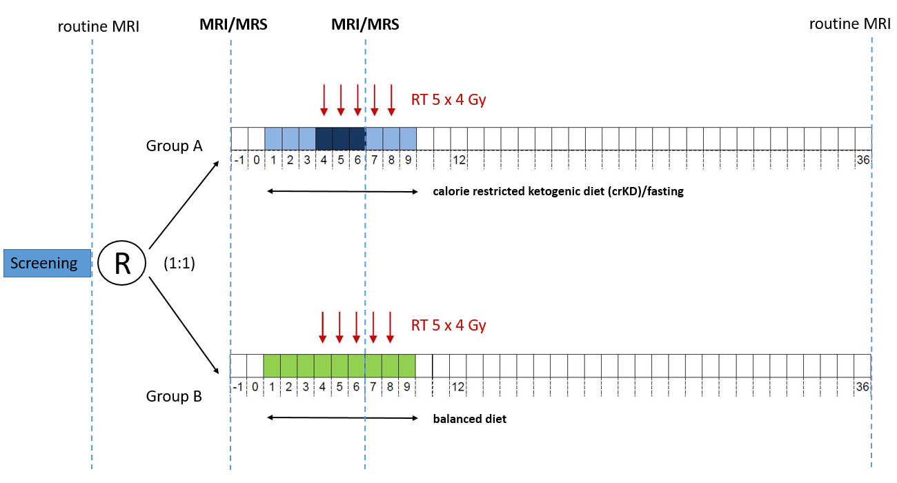
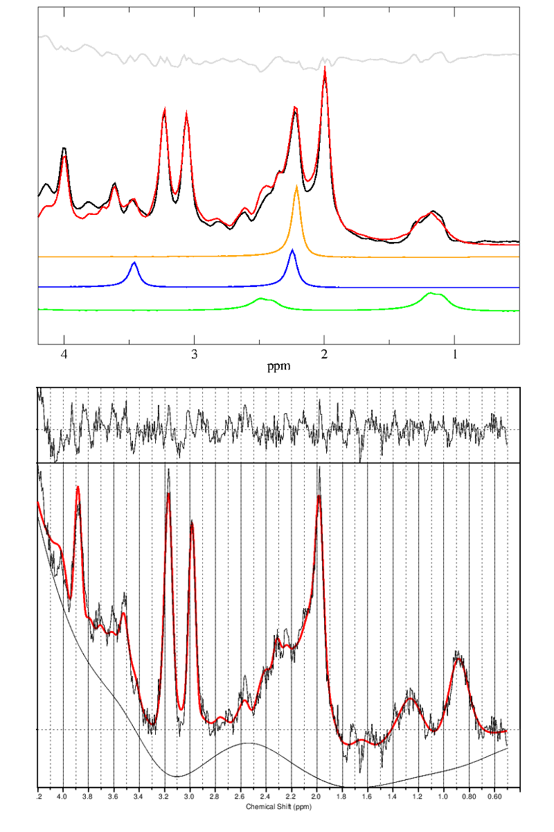
This report is based on a prospective multicentric, non-blinded, randomized study, including an extended MRS protocol (1H decoupled 31P MRSI with 3D CSI and 2D 1H CSI, Fig.1) at a 3T clinical scanner. Study population consisted of patients with recurrent glioblastoma and indication for re-irradiation therapy. 50 patients enrolled at all centers were randomized (1:1) in two groups: Group A keeping a ketogenic diet with calorie restriction (21-23 kcal/kg/day) for nine days, including three days of fasting (0 kcal/day) and group B keeping a balanced diet over the same period. RT was performed on day 4-8 (5 x 4 Gy). 32 patients received MRS examination at baseline (day-1) and 23/32 on day 6 (Fig.3). Registration to 3D-anatomical data was performed with an in-house software tool scripted in Matlab. Voxels were selected from the area of recurrent tumor and on the contralateral hemisphere in NAWM (control). Analysis of proton data was performed using LCModel and phosphorous data were analyzed with jMRUI (AMARES) (5, 6). All spectra were inspected for quality. The basis set for LCModel including 3-hydroxybutyrate (βOHB), acetone (Ac) and acetoacetate (AcAc) was simulated using the NMRScope-B plugin implemented in jMRUI (7) and tested on phantom data (Fig.5). Results were considered to be significant at p<0.05 using a non-parametric t-test.Results
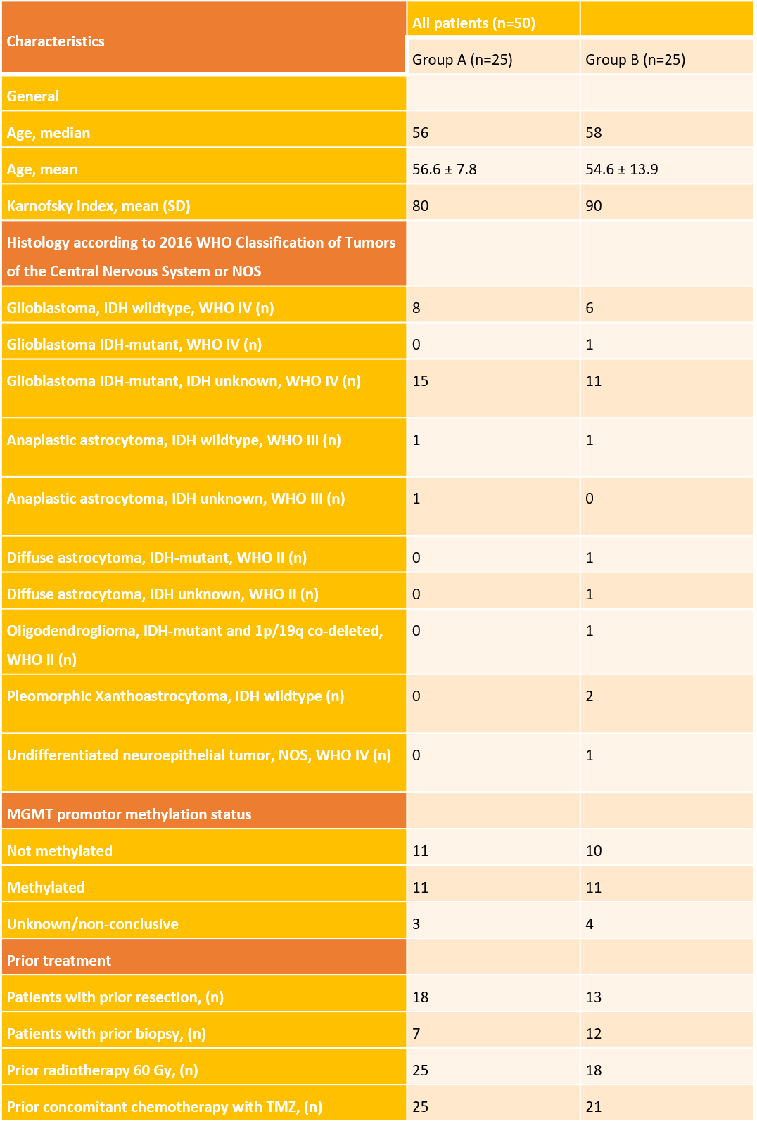
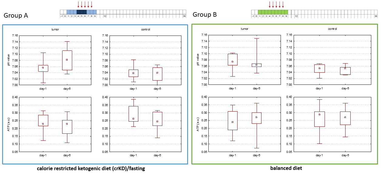
Patient characteristics and treatment are summarized in Fig.3. 17/25 patients in group A achieved a serum ketone level of >0.5 mmol/l at day 6 during crKD/fasting (dropout: 4 patients). Overall dietary requirements were well tolerated. There was a significant difference in serum ketone levels at day 6 between both groups. KB were detected within tumor tissue in two patients of group A at day 6 with an estimated standard deviation (%SD) ≤30% (LCModel). Both patients displayed an acetone signal at 2.22 ppm which was quantified. One of these patients correspondingly showed a high serum ketone level of 4.5 mmol/l. As expected, for both groups, pHi was significantly lower in control voxels than in tumor voxels (day 6 Group A: p=0.007, Group B: p=0.018). Neither pHi nor ATP levels showed significant changes in control voxels of either group. Group A exhibited a significant increase in pHi in tumor voxels comparing baseline levels to day 6 during crKD/fasting (p=0.027), while there were no changes reported for Group B. In both groups ATP levels were stable (Fig.4). With eight patients still censored overall survival (OS) and progression free survival analysis is pending.Discussion
Even with elevated ketone serum levels in most patients in group A, MRS-detection of Acn was only seen within tumor tissue in two patients. A lack in correlation to serum ketone levels and late appearance of brain changes has been previously reported, even though we were able to detect intracerebral KB as early as day 6 of crKD/fasting (8) The fact that Acn was detected in tumor tissue only might be due to impaired BBB (9). Recently, the hypothesis that brain tumors are metabolically inflexible has been contradicted in two rat glioma models with upregulation of the ketone-body monocarboxylate transporter, facilitating uptake and oxidation of KB in tumor cells (10). The proposed metabolic flexibility might lead to KB oxidation and a shift towards the somewhat impaired mitochondria. A possible result could be improved energy resources for active transport of H+-equivalents, resulting in an even more alkaline pHi. This might support tumor metabolism and lead to a lack of improvement in OS for Group A.Conclusion
We were able to show evidence of intratumoral Acn using 1H-MRS in some patients with malignant glioma at day 6 of crKD/fasting. Changes in pHi and ATP during crKD/fasting will remain subject to preclinical studies.Acknowledgements
No acknowledgement found.References
1. Kiebish MA, Han X, Cheng H, Seyfried TN. In vitro growth environment produces lipidomic and electron transport chain abnormalities in mitochondria from non-tumorigenic astrocytes and brain tumours. ASN Neuro. 2009;1(3). doi:10.1042/AN20090011.
2. Maurer GD, Brucker DP, Bähr O, et al. Differential utilization of ketone bodies by neurons and glioma cell lines: a rationale for ketogenic diet as experimental glioma therapy. BMC Cancer. 2011;11:315. doi:10.1186/1471-2407-11-315.
3. Oudard S, Boitier E, Miccoli L, Rousset S, Dutrillaux B, Poupon MF. Gliomas are driven by glycolysis: putative roles of hexokinase, oxidative phosphorylation and mitochondrial ultrastructure. Anticancer Res. 1997;17(3C):1903-1911.
4. Marathe K, McVicar N, Li A, Bellyou M, Meakin S, Bartha R. Topiramate induces acute intracellular acidification in glioblastoma. J Neurooncol. 2016;130(3):465-472. doi:10.1007/s11060-016-2258-y.
5. Naressi A, Couturier C, Castang I, Beer R de, Graveron-Demilly D. Java-based graphical user interface for MRUI, a software package for quantitation of in vivo/medical magnetic resonance spectroscopy signals. Comput Biol Med. 2001;31(4):269-286.
6. Vanhamme, van den Boogaart A, van Huffel S. Improved method for accurate and efficient quantification of MRS data with use of prior knowledge. J Magn Reson. 1997;129(1):35-43.
7. Starcuk Z, Strbak O, Starcukova J, Graveron-Demilly D. Simulation of steady state free precession acquisition mode in coupled spin systems for fast MR spectroscopic imaging. In: 2008 IEEE International Workshop on Imaging Systems and Techniques: IEEE; 2008 - 2008:302-306.
8. Artzi M, Liberman G, Vaisman N, et al. Changes in cerebral metabolism during ketogenic diet in patients with primary brain tumors: 1H-MRS study. J Neurooncol. 2017;132(2):267-275. doi:10.1007/s11060-016-2364-x.
9. Musa-Veloso K. Non-invasive detection of ketosis and its application in refractory epilepsy. Prostaglandins Leukot Essent Fatty Acids. 2004;70(3):329-335. doi:10.1016/j.plefa.2003.08.025.
10. Feyter HM de, Behar KL, Rao JU, et al. A ketogenic diet increases transport and oxidation of ketone bodies in RG2 and 9L gliomas without affecting tumor growth. Neuro-oncology. 2016;18(8):1079-1087. doi:10.1093/neuonc/now088.
Figures