2900
Eye-movements and white matter predict emotional control in children treated for brain tumorsIska Moxon-Emre1,2,3, Michael D Taylor1,2, Adeoye Oyefiade 1,2, Eric Bouffet1,2, Suzanne Laughlin1,2, Jovanka Skocic 1, Cynthia B de Medeiros 1, and Donald J Mabbott1,2
1The Hospital for Sick Children, Toronto, ON, Canada, 2The University of Toronto, Toronto, ON, Canada, 3Pediatric Oncology Group of Ontario, Toronto, ON, Canada
Synopsis
Children who survive a brain tumor diagnosis often suffer from emotional difficulties that decrease their quality of life. We monitored eye-movements during the control of attention to emotional faces to measure emotion regulation. Brain tumor survivors had difficulty regulating their initial attention away from emotional faces, and those who exhibited poor emotion regulation displayed the least emotional control in daily life. White matter of the splenium of the corpus callosum predicted emotion regulation. Our findings may improve the identification of children at risk for poor functional outcomes, and suggest the splenium as a candidate neuroanatomic substrate of emotion regulation.
Introduction
Children treated for brain tumors, including those that arise in the posterior fossa (PF), often experience social and emotional dysfunction that impair their functioning and decrease their quality of life. There are no biomarkers to detect and quantify this impairment, and the neuroanatomic basis is currently unknown. To understand emotional functioning and to identify neuroanatomical predictors of emotional dysfunction in children treated for brain tumors, multi-method and multi-informant measures are necessary. Here, for the first time in children treated for PF tumors, we combine an objective eye-tracking measure with a standardized measure to evaluate emotion regulation, and we evaluate the associations between emotion regulation and WM organization using Diffusion Tensor Imaging (DTI).Methods
Fifty-four children participated in this study; 36 children treated for PF tumors (17 patients treated with surgery with or without chemotherapy, and 19 patients treated with surgery, chemotherapy and radiation) at the Hospital for Sick Children (SickKids; Toronto, Canada) and 18 healthy control children. Participants completed two versions of an eye-tracking task: 1. Baseline (free-viewing) condition: Images of emotional/neutral face pairs were presented side-by-side, and participants were instructed to look at the faces freely. 2. Regulate (directed-viewing) condition: A second set of emotional/neutral pairs were presented side-by-side, and participants were instructed to look at the non-emotional face only. An emotion regulation score was calculated by subtracting the time to first fixation to the target (i.e., neutral) from the non-target (i.e., emotional) face. Eye movements were recorded throughout all tasks using a SR Research Ltd. Eyelink 1000 plus eye-tracking desktop monocular system. A sampling rate of 500 Hz and a spatial resolution of 0.01° was used. To evaluate a child’s ability to appropriately modulate their emotional responses, the ‘emotional control’ scale from the parent-report Behavior Rating Inventory of Executive Function (BRIEF) questionnaire was used. Magnetic Resonance Imaging (MRI) was performed using a Siemens 3T whole-body MRI scanner (Prisma fit) with a 12-channel head coil. Imaging included a T1 AX 3D MPRAGE Grappa 2 protocol (T1=900ms, TE/TR=3.83/2300ms, 160 contiguous axial slices, flip angle=9°, 256x224 matrix, FOV=256x224mm, voxel size=1mm ISO) and diffusion-weighted single shot spin echo DTI sequence with EPI readout (30 directions, b=1000s/mm2, TE/TR=90/9000ms, 70 contiguous axial slices, flip angle=90°, 122x122 matrix interpolated to 244x244, FOV=244x244mm, voxel size=2mm ISO, interpolated to 1x1x2mm). First, time to first fixation, an eye-tracking measure of attentional capture, was compared between control and patient groups during both conditions, using mixed design ANOVAs. Second, the eye-tracking emotion regulation score was correlated with the emotional control score. Third, a whole brain voxel-based analysis was conducted to assess if FA and RD in any voxels throughout the brain correlated with our eye-tracking emotion regulation measure, in controls and patients considered separately.Results
In the baseline condition, the emotional face captured attention across all groups; there was main effect of emotion for the time to first fixation (p<0.0001). In the regulate condition, a significant emotion by group interaction (p=0.007) revealed that only healthy controls had a shorter time to first fixation to the neutral face compared to the emotional face (p=0.0007) (Fig 1a), indicating that only controls were able to override the attentional capture of emotional faces when instructed to. Across all children, the emotional control score was positively correlated with the eye-tracking emotion regulation score (r=0.29, p=0.045). In patients, there were many clusters of voxels throughout the brain where FA was positively correlated (Fig 1b), and RD was negatively correlated with the eye-tracking emotion regulation score (higher score = worse regulation) (all p<0.05), located mostly in the splenium. In healthy controls, there were no voxels where RD or FA correlated with the eye-tracking emotion regulation score (all p>0.05).Discussion and Conclusion
Our findings demonstrate that patients treated for brain tumors have difficultly regulating the attentional capture of emotional faces. We also demonstrate that better microstructural organization of white matter in the splenium predicts worse emotion regulation in patients. Across all groups, children who have difficultly regulating the attentional capture of emotional faces during their earliest visual response display worse emotional control in daily life. That our novel eye-tracking based biomarker predicts daily function, suggest a simple early oculomotor measure of attention can inform complex human behavior. Our findings may help identify children treated for PF tumors that are at risk for poor functional outcomes, and also suggests the splenium of the corpus callosum as a candidate neuroanatomic substrate of emotion regulation.Acknowledgements
This work was supported by the Canadian Institute of Health Research (CIHR) and the Pediatric Oncology Group of Ontario (POGO).References
No reference found.Figures
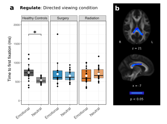
a. Boxplots showing all data points with the mean (white diamond) and
median (black line) for the time to first fixation to the emotional face vs.
neutral face in the regulate condition, *p = 0.0007; Bonferroni corrected
pairwise comparison. b. Controlling
for age, voxels where FA (blue) was positively correlated, with the
eye-tracking emotion regulation score in children treated for brain tumors
(higher scores = worse regulation).