2896
Measurement of peripheral nerve perfusion using FAIR PRESS1Department of Radiology, German Cancer Research Center, Heidelberg, Germany, 2Department of Neuroradiology, Heidelberg University Hospital, Heidelberg, Germany
Synopsis
Peripheral neuropathy in diabetes is a common and poorly understood disease; previous methods to evaluate nerve-associated microvascular angiopathy are contrast-based and exclude patients with severe nephropathies or allergies to contrast agents. We adapt a combined a flow-sensitive alternating inversion recovery (FAIR)-sequence with a single-voxel readout such as Point RESolved Spectroscopy (PRESS) to measure peripheral nerve perfusion in healthy subjects and diabetes patients. Our preliminary results suggest that diabetes patients have a lower nerve/muscle perfusion ratio than non-diabetic subjects.
INTRODUCTION
While distal diabetic neuropathy is a frequent and yet poorly understood complication of diabetes, neural changes are associated with changes in lipid metabolism and microvascular angiopathy1. Previous measurements of neural perfusion, however, use contrast-enhanced imaging that excludes patients with severe nephropathies or allergies to contrast agents. By combining a flow-sensitive alternating inversion recovery (FAIR)-sequence with a single-voxel readout, such as Point RESolved Spectroscopy (PRESS), non-invasive perfusion measurements of tissues with low signal noise ratio is possible. This technique has been applied to access white matter perfusion2,3 and rat skeletal muscle perfusion4. In this study, we adapt the FAIR PRESS sequence to measure peripheral nerve perfusion in healthy subjects and diabetes patients. The perfusion of peripheral nerve is compared to that of skeletal muscle.METHODS
6
subjects (4 healthy; 2 diabetes) were scanned using a 15-channel knee
transmit/receive RF coil on a 3T Trio Siemens scanner (Siemens Healthcare, Erlangen, Germany).
Sequence parameters of FAIR PRESS with QUIPSS II5 were as follows:
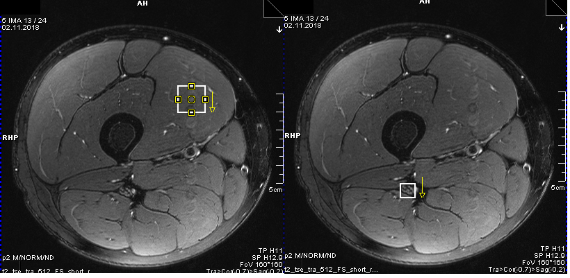
TE/TR=30/2300 ms, TI1/TI=800/1500 ms, voxel size of muscle=15×15×15
mm3, voxel size of nerve=8×8×8 mm3, spectral
bandwidth=2000 Hz, measurements=60 (30 control-label pairs). The placement of
muscle and nerve voxels was based on a T2 measurement (Figure 1).
Perfusion analysis was performed using in-house developed MATLAB code
(MathWorks, Natick, MA). The free induction decays (FIDs) of each acquisition
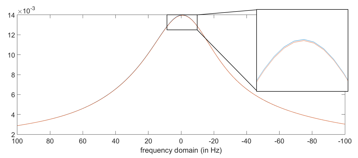
of FAIR PRESS were truncated, zero filled, filtered with an apodization filter
of 15 Hz, phase corrected, and Fourier transformed. The data were then
combined by adding the root of the sum of squares of the individual signals
from the different coils. The central 100 points of the water line peak were
used for perfusion analysis (Figure 2). Perfusion quantification was performed
as in literature6.RESULTS
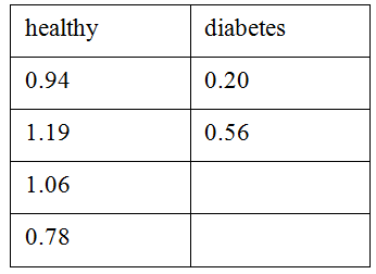
Perfusion ratios between nerve and muscle from all subjects are presented in Table 1. Relative lower nerve perfusion was found in diabetes patients. However, the sample size is still too small for a fair comparison between healthy subjects and diabetes patients.CONCLUSION
This
study demonstrates the feasibility of perfusion measurement of peripheral nerve
using a FAIR PRESS sequence. While still preliminary, a comparison between
diabetic and healthy subjects shows differences in neural perfusion. The
sequence may thus aid in the evaluation of the extent and correlation of
nerve-associated angiopathy with neural lesions.Acknowledgements
No acknowledgement found.References
1. Jende JME, Groener JB, Oikonomou D, et al. Diabetic neuropathy differs between type 1 and type 2 diabetes: Insights from magnetic resonance neurography. Ann Neurol 2018;83(3):588-598.
2. Pohmann R. Accurate, localized quantification of white matter perfusion with single-voxel ASL. Magn Reson Med 2010;64(4):1109-1113.
3. Zhang X, Ronen I, Kan HE, et al. Time-efficient measurement of multi-phase arterial spin labeling MR signal in white matter. NMR Biomed 2016;29(11):1519-1525.
4. Pohmann R, Kunnecke B, Fingerle J, et al. Fast perfusion measurements in rat skeletal muscle at rest and during exercise with single-voxel FAIR (flow-sensitive alternating inversion recovery). Magnet Reson Med 2006;55(1):108-115.
5. Wong EC, Buxton RB, Frank LR. Quantitative imaging of perfusion using a single subtraction (QUIPSS and QUIPSS II). Magnet Reson Med 1998;39(5):702-708.
6. Alsop DC, Detre JA, Golay X, et al. Recommended Implementation of Arterial Spin-Labeled Perfusion MRI for Clinical Applications: A Consensus of the ISMRM Perfusion Study Group and the European Consortium for ASL in Dementia. Magnet Reson Med 2015;73(1):102-116.
Figures