2895
T1, T2, and STIR-weighted spine MRI using spiral SE/TSE techniques1Department of Neuroradiology, Barrow Neurological Institute, Phoenix, AZ, United States, 2Department of Radiology, Mayo Clinic, Rochester, MN, United States
Synopsis
Spine MRI constitutes a significant portion of neuro exams but is difficult in the clinical practice. The challenges include inhomogeneous field, motion and pulsation induced artifacts, long scan times, high SAR, etc. To overcome these drawbacks, in this work we explore the fast spiral sampling approach with SE and TSE sequences to provide rapid T1 and T2 weighted water-fat imaging, as well as STIR imaging. The feasibility is demonstrated with sagittal c-spine scans on volunteers.
INTRODUCTION
Spine MRI constitutes a significant portion of clinical neuro exams, and often acquires T1, T2, and/or STIR (short TI inversion recovery) weighted images using the TSE sequence. The common issues of TSE spine imaging include inhomogeneous field, motion and pulsation induced artifacts, long scan times, high specific absorption rate (SAR), etc.1 For certain spine applications, water-fat separated images may offer valuable supplemental information. However, adding Dixon water-fat imaging2 in conventional TSE spine scans either significantly increases the scan time, or incurs signal to noise ratio (SNR) loss if parallel imaging is employed. Compared to Cartesian acquisition, spiral sampling provides efficient data acquisition and insensitivity to motion and flow related artifacts. Recently spiral SE and TSE have regained attention for T1 and T2 weighted brain imaging.3,4 Nonetheless, it is still challenging to image the spine with spiral acquisition in the sagittal plane due to the anatomical shape and inhomogeneous field. In this project, we explore the spiral SE and TSE sequences for T1, T2, and STIR weighted imaging and demonstrate the feasibility with volunteer c-spine scans.METHODS
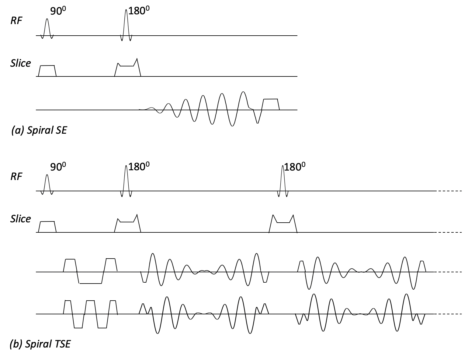
The spiral protocols for sagittal spine imaging include T1 weighted imaging using a spiral SE sequence3, and T2 and STIR weighted imaging with a spiral TSE sequence4. The pulse sequences are illustrated in Fig. 1. An anisotropic resolution is implemented in the spiral sequence to match the Cartesian scans, which results in an anisotropic field of view (FOV).5 T1 and T2 data are acquired with 2 TE shifts for Dixon-based water-fat separation. The spiral TSE technique uses a spiral-in/out trajectory to improve efficiency as well as robustness to off-resonance artifacts, and a double-encoding acquisition strategy along with additional signal demodulation in reconstruction to minimize T2-decay induced artifacts.4 A pre-scan is performed prior to all spiral scans to generate a B0 field map that is used in the spiral data reconstruction for deblurring and/or water-fat separation6.
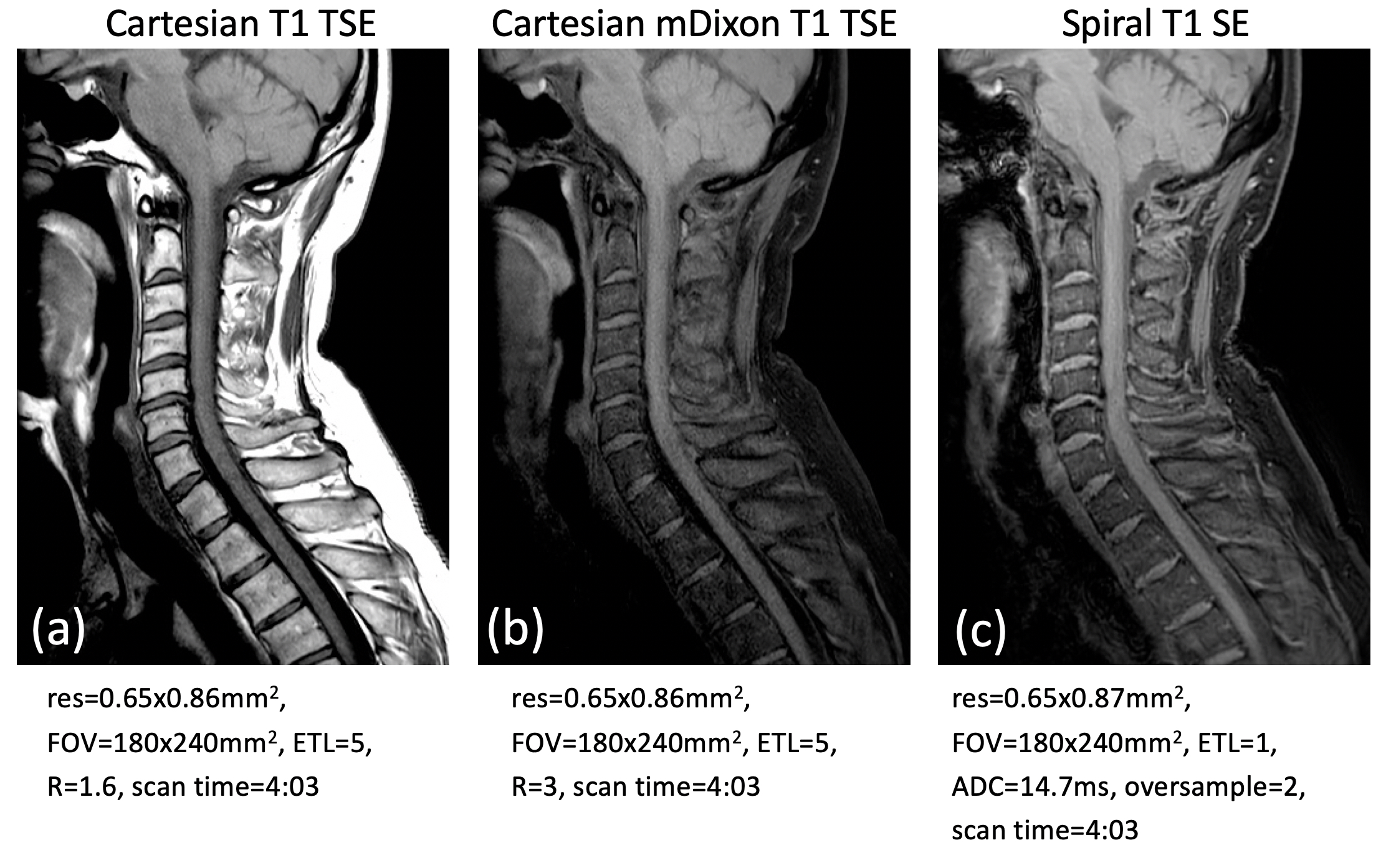
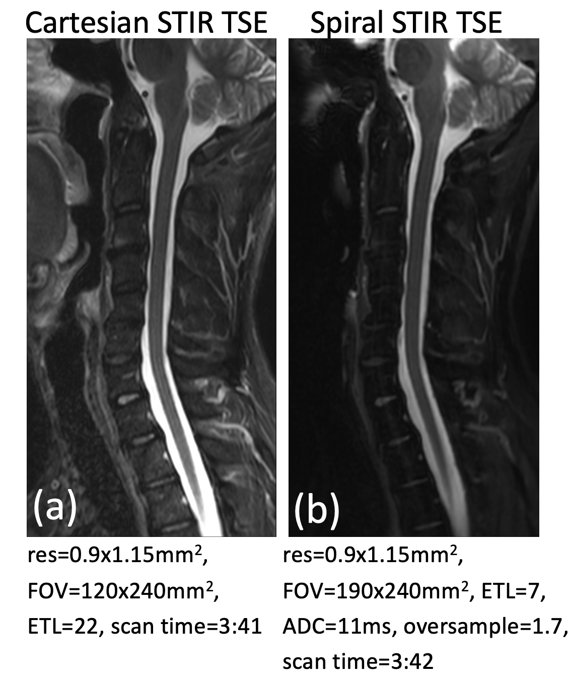
Volunteers were scanned on a 3T Philips Ingenia scanner using a Head NV coil. T1, T2, and STIR data were acquired with Cartesian TSE and spiral SE/TSE. T1 and T2 weighted Cartesian data were acquired with and without Dixon imaging. Parameters such as sampling time (ADC) and echo train length (ETL) are adjusted in the spiral sequences to achieve comparable TE/contrast as the Cartesian scans. To match the scan time, the SENSE reduction factor R in the Cartesian scans and the oversampling factor in the spiral scans are also adjusted correspondingly. The parameters are listed below each image in the figures in the Results section.
RESULTS
The results are shown in Figs. 2~4. For data acquired with Dixon imaging, only the water images are demonstrated. Fig. 2 compares the T1 weighted images acquired with (a) Cartesian TSE, (b) Cartesian mDixon TSE, and (c) spiral SE. Fig. 3 illustrates the T2 weighted images from Cartesian TSE (a) without and (b) with mDixon, and (c) spiral TSE. Fig. 4 shows the STIR images from (a) Cartesian and (b) spiral TSE. Without mDixon, Cartesian TSE generates good SNR in both T1 and T2 images (Figs. 2a and 3a). However, the SNR is reduced in Cartesian mDixon TSE images (Figs. 2b and 3b). With spiral acquisition, good SNR and image quality are achieved in both T1 and T2 weighted data (Figs. 2c and 3c), as well as STIR images (Fig. 4b).DISCUSSION
As shown in the Results section, the spiral techniques provide the capability of water-fat imaging while preserving high SNR. Also reflected by the short ETL in the spiral scans is the significantly reduced SAR. The SENSE reduction factor in the Cartesian scans and the oversampling factor in the spiral scans also indicate the fast speed of the spiral acquisition, which can be used to further reduce the scan time. In summary, the spiral SE/TSE techniques can be adapted for the spine anatomy and provides a potential tool for robust spine MRI.Acknowledgements
This work was partially funded by Philips Healthcare.References
- Stroman PW, Wheeler-Kingshott C, Bacon M, et al. The Current State-of-the-Art of Spinal Cord Imaging: Methods. Neuroimage. 2014;84:1070-1081.
- Dixon WT. Simple Proton Spectroscopic Imaging. Radiology. 1984;153:189-194.
- Li Z, Hu HH, Miller JH, et al. A Spiral Spin-Echo MR Imaging Technique for Improved Flow Artifact Suppression in T1-Weighted Postcontrast Brain Imaging: A Comparison with Cartesian Turbo Spin-Echo. Am J Neuroradiol. 2016;37:642-647.
- Li Z, Karis JP, Pipe JG. A 2D Spiral Turbo Spin-Echo Technique. Magn Reson Med. 2018; 80:1989-1996.
- King KF. Spiral Scanning with Anisotropic Field of View. Magn Reson Med. 1998;39:448-456.
- Wang D, Zwart NR, Li Z, et al. Analytical Three-Point Dixon Method: With Applications for Spiral Water-Fat Imaging. Magn Reson Med. 2016;75;627-638.
Figures