2894
Super multi-directional DTI for lumbar nerve roots using MultiBans SENSE1Eastern Chiba Medical Center, Chiba, Japan, 2Graduate School of Medical Sciences, Kanazawa University, Ishikawa, Japan, 3Philips Japan, Tokyo, Japan
Synopsis
An increasing the number of MPG directions causes prolongation of scan time. Recently, MultiBand SENSE (MB-SENSE) has been developed to allow the simultaneous acquisition of multiple slices and it enabled to shorten the scan time. Our purpose in this study was to investigate whether super multi-directional DTI using MB-SENSE is more useful for evaluation of lumbar nerve disorders than conventional method. An increasing the number of MPG directions improved its continuity and accuracy in tractography and tended to decrease the standard deviation of FA values.
Purpose
Diffusion tensor imaging (DTI) based on a single-shot echo planer imaging (EPI-DTI) is an established method that has been used for evaluation of lumbar nerve disorders in previous studies1. There has been reported that the nerve continuity in tractography can be improved by increasing the number of Motion Probing Gradient (MPG) directions2-3, but an increasing the number of MPG directions causes prolongation of scan time. Thus, it is challenging in routine clinical MRI so far. Recently, MultiBand SENSE (MB-SENSE) has been developed to allow the simultaneous acquisition of multiple slicesand it enabled to shorten the scan time4. Our purpose in this study was to investigate whether super multi-directional DTI using MB-SENSE is more useful for evaluation of lumbar nerve disorders than conventional method.Method
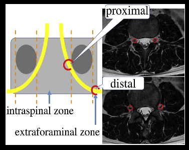
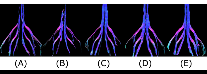
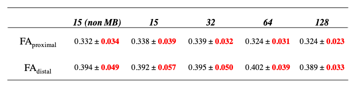
All subjects were examined by use of a 3.0 T whole-body clinical system (Ingenia CX, Philips Healthcare) with a dedicated 32-channel flexible torso coil. DTI imaging was performed on the lumbar nerve roots (L4to S1) of 10 healthy volunteers. The imaging parameters were the conventional method with 15 directions and the number of MPG directions (15, 32, 64, 128) using MB technique. b-factor was 800 s/mm2. For tractography and measurement of FA values,ROIs were placed both proximally and distally to the lumbar foraminal zone at two levels on the L4 to S1 nerve roots (Fig.1). We examined visual evaluation (continuity, accuracy) and quantitative evaluation (FA value) in tractographyunder each imaging parameters.Result
Regarding the subjective visual evaluation, the number of MPG directions improved its continuity and accuracyin tractography as increased MPG directions(Fig.2). Regarding the subjective quantitativeevaluation, the number of MPG directions tended to decrease the standard deviation of FA values as increased MPG directions(Fig.3). This is because a large number of MPG directions can determine the direction of water molecule diffusion more accurately. Although further clinical evaluation is needed, 128 MPG directionsis the optimal value for improving the quality of DTI and its Tractography.Conclution
The super multi-directional DTI (128 MPG directions) using MB-SENSE clearly improves the visualization capability of lumbar nerve and increases the accuracy of FA values within the clinically feasible acquisition time.Acknowledgements
No acknowledgement found.References
[1] Balbi V, Budzik JF, Duhamel A, Bera-Louville A, LeThuc V, Cotton A. Tractography of lumbar nerve roots: initial results. Eur Radiol 2011;21:1153–9
[2] Takayuki Sakai, et al. Distortion-free diffusion tensor imaging for evaluation of lumbar nerve roots: Utility of direct coronal single-shot turbo spin-echo diffusion sequence. Magnetic Resonance Imaging 2018;49:78–85
[3] Akiyoshi Hamaguchi, Shuuichi Kodera. Influence Exerted by MPG-directions on Diffusion Tensor Imaging(DTI). Jpn.J. Radiol Technol 2009;7(65):913-920
[4] David A Feinberg, Kawin Setsompop. Ultra-fast MRI of the human brain with simultaneous multi-slice imaging. J Magn Reson Imaging 2013;229:90-100
Figures