2876
Structural Assessment of Injured Spinal Cord Using the Spherical Mean Technique and Diffusion Tensor Imaging1Vanderbilt University Institute of Imaging Science, Vanderbilt University Medical Center, Nashville, TN, United States, 2Department of Radiology and Radiological Sciences, Vanderbilt University Medical Center, Nashville, TN, United States, 3Department of Biomedical Engineering, Vanderbilt University, Nashville, TN, United States
Synopsis
High-resolution spherical mean technique (SMT) MRI may provide specific diffusion measures to detect and characterize structural connectivity before and after spinal cord injury and during repair. This study aims to systematically evaluate the SMT-derived diffusion parameters and compare them to conventional DTI-derived diffusion parameters in assessing injury-associated changes in spinal cords of squirrel monkeys. The main goal is to optimize a rapid and robust high-resolution SMT imaging protocol for spinal cord of monkeys at high field. In addition, the sensitivity of SMT and DTI measures in detecting axonal damage in the dorsal pathway has been evaluated.
Purpose
This study aims to systematically
evaluate the feasibility and precision of diffusion measurements based on the
spherical mean technique (SMT) for assessing microstructural properties of the spinal
cords of non-human primates. This study also aims to characterize changes in different
diffusion measures in individual subjects, both regionally and longitudinally,
in order to evaluate their sensitivity for detecting tissue structural change
after cervical spinal cord injury (SCI).Methods
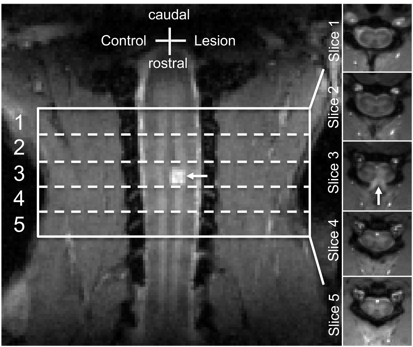
MRI scans were performed on isoflurane-anesthetized squirrel monkeys at 9.4T, before and after a unilateral section (~2 mm) of the dorsal column white matter tract (Fig. 1).1 The spin-echo diffusion sequence used an echo planar imaging readout (TR/TE = 3000/33 ms, 4 shots, resolution = 0.333x0.333 mm2, slice thickness = 3 mm, 5 slices). For diffusion imaging, three b-shells were acquired with b values at 750, 1000, and 2000 s/mm2 respectively, sampling 30 directions. Three non-diffusion weighted scans were acquired, one at the beginning of each b-value shell acquisition. SMT data were acquired from the cervical spinal cords of seven normal and five spinal cord injured monkeys. SMT derived the apparent axonal volume fraction Vax and intrinsic axonal diffusivity Dax.2 The extra‐axonal transverse diffusivity Dex was estimated as a function of Vax and Dax. The influences of variations in the acquisition scheme on SMT were evaluated. Conventional DTI parameters including the fractional anisotropy (FA), axial diffusivity (AD), radial diffusivity (RD), and mean diffusivity (MD) were also quantified for comparison. Regional correlations between diffusion measures were calculated using the Pearson correlation function. The significance of group regional differences was evaluated using Student’s t-tests. P < 0.05 was considered statistically significant.Results
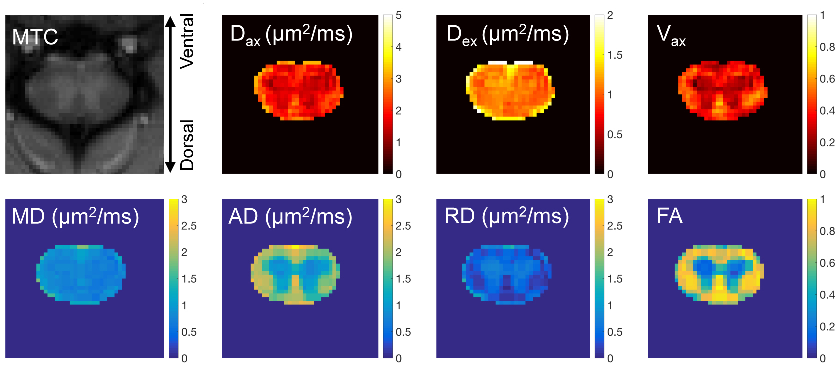
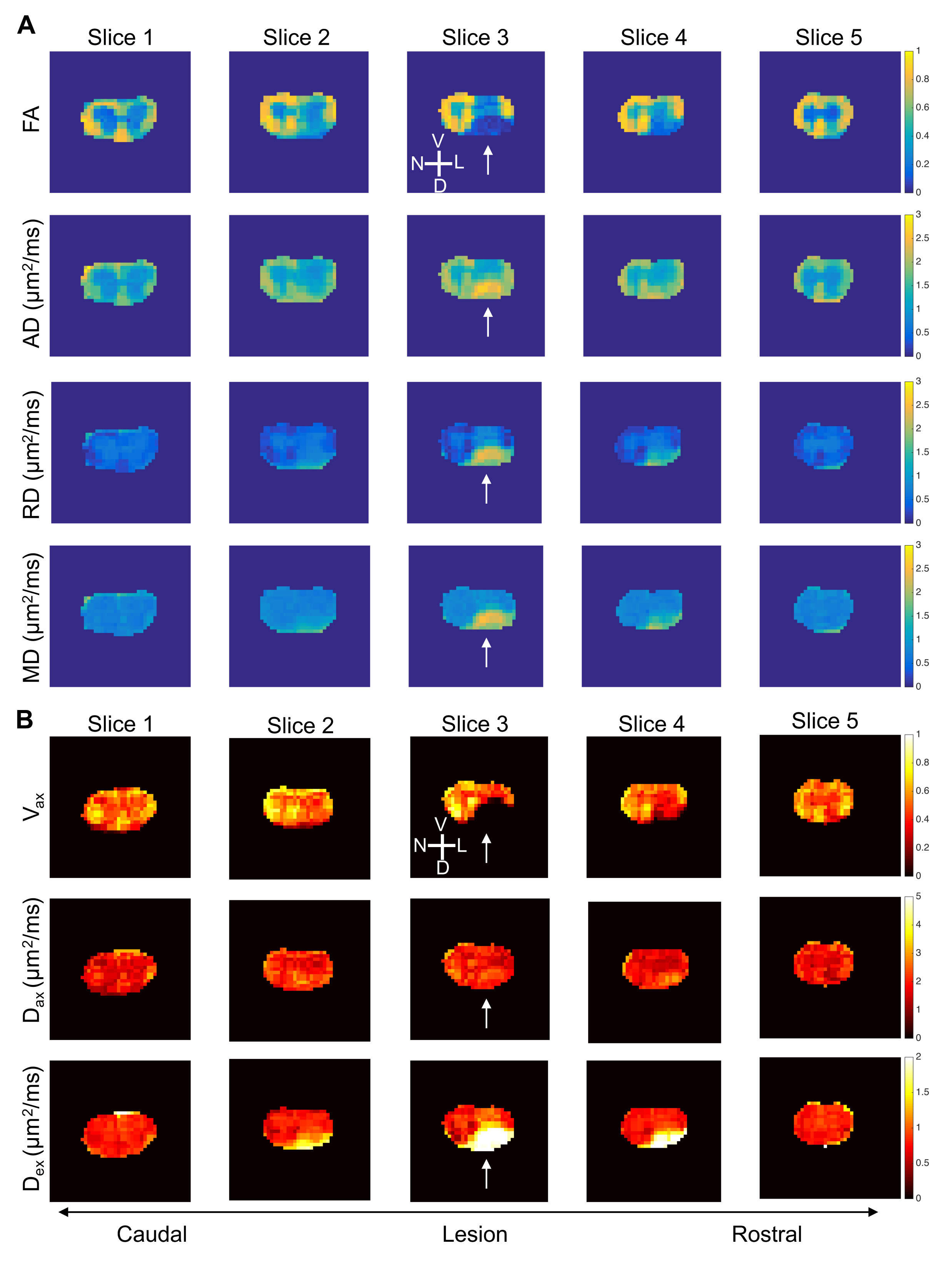
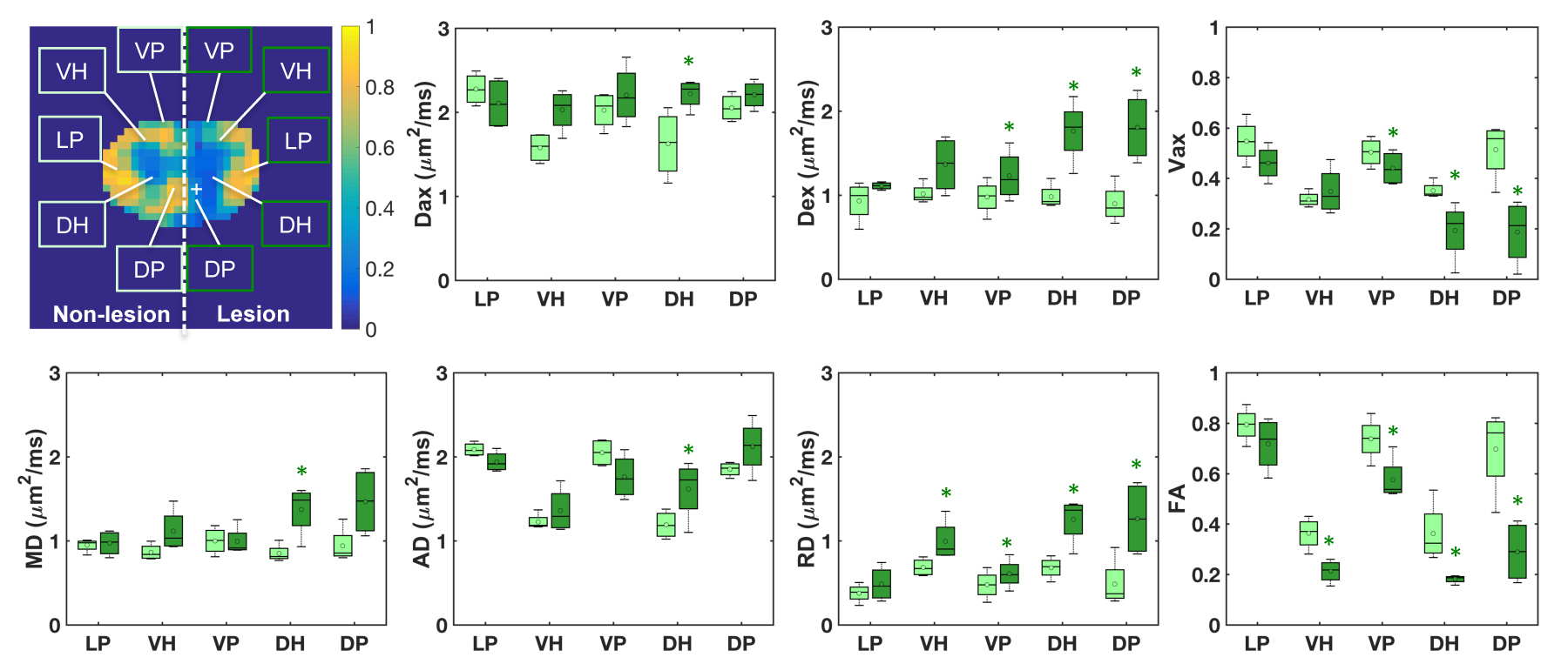
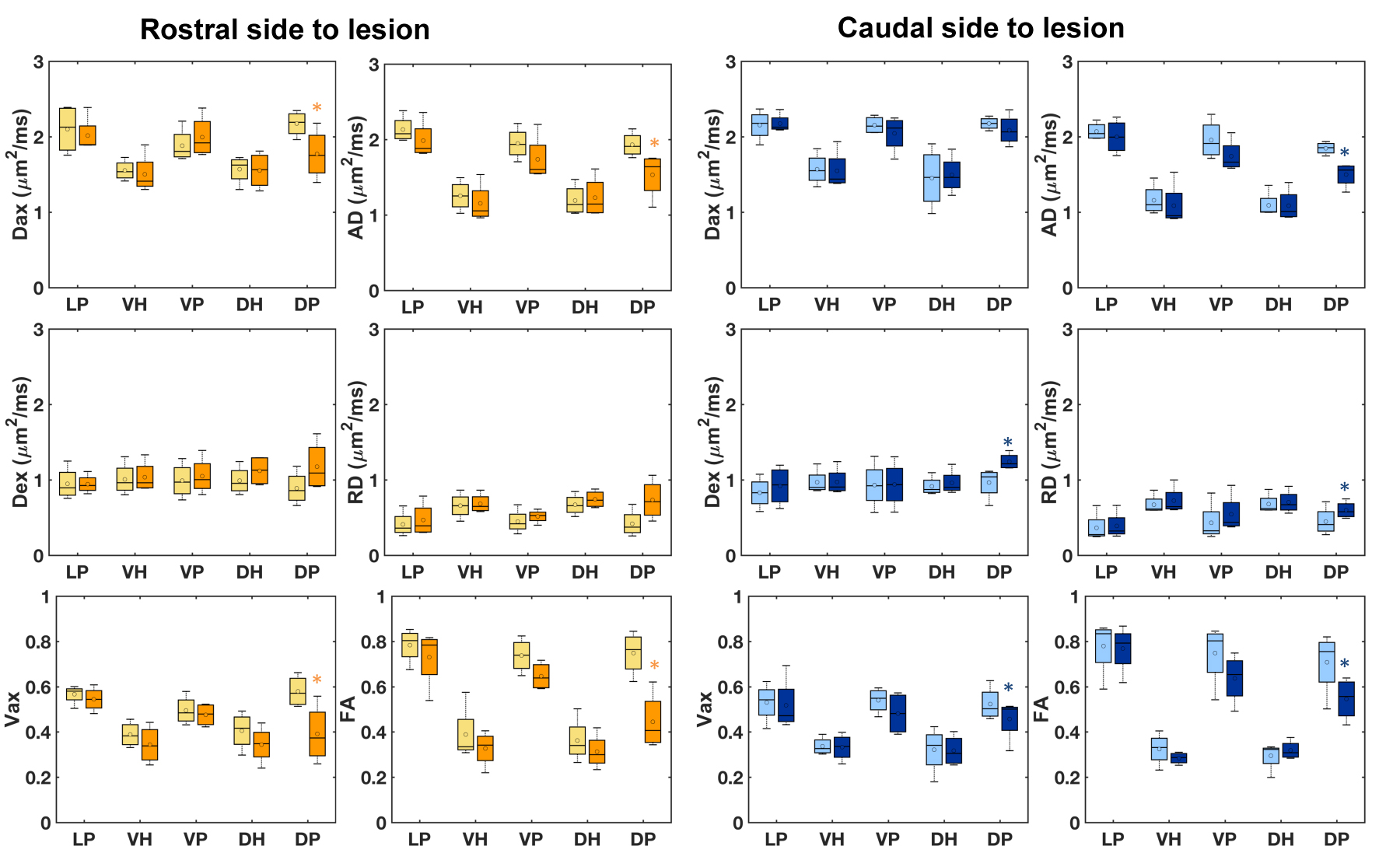
DTI and SMT measurements delineated normal white matter (WM) and gray matter (GM) in the cervical spinal cord of monkey (Fig. 2) with good contrast. Regions of WM (LPN, VPN and DPN) showed significantly higher Vax and Dax than GM (VHN and DHN), and conventional DTI parameters FA and AD showed similar regional trends. The measured Dex of GM was slightly higher than that of WM, while RD of GM was significantly higher than that of WM. The SMT-derived measures Vax and Dax were strongly correlated with each other, and they also showed high correlation with DTI measures FA and AD. Although DTI measures showed higher precision across slices than SMT measures at the same SNR level, it is of note that a twofold reduction in acquisition yielded comparable accuracy with SMT. All the DTI and SMT measures detected unilateral changes at the site of injury, especially in the dorsal pathway (Figs. 3-4). A significant decrease in Vax was also observed in rostral and caudal regions next to the lesion site (p < 0.05), while FA decreased. Although both Dax and Dex increased at the lesion site, Dex showed higher unilateral contrast than Dax compared to the non-lesion control side twelve days after spinal cord injury. In this representative injured subject, the rostral region to the lesion showed a more severe damage than the caudal region (Fig. 3). This is also seen in the statistical results across subjects (Fig. 5). It is notable that Dax and AD increased at the lesion/cyst but decreased in the tissues of the dorsal pathway rostral or caudal to the lesion site (Figs. 4-5). SMT-derived Vax was sensitive for detecting axonal damage and recovery in the dorsal column pathway after unilateral dorsal column lesion, comparable to FA (Figs. 3-5). Longitudinally, evident recovery from injury is revealed by changes of these diffusion parameters.Conclusion
Diffusion imaging can provide an efficient and sensitive means to detect and characterize axonal damage after injury and monitor its recovery over time. SMT modelling provides a robust and alternative method to DTI analysis for characterization of the damage of spinal cord after injury, independent of axonal orientation.Acknowledgements
We thank Mrs. Chaohui Tang and Mr. Fuxue Xin of the Vanderbilt University Institute of Imaging Science for their assistance in animal preparation and care in MRI data collection. We also thank Mr. Ken Wilkens and Dr. Xinqiang Yan for customizing coils for cervical spinal cord imaging. This study is supported by DOD grant W81XWH-17-1-0304, and NIH grant NS092961.References
1. Wang F, Zu ZL, Wu RQ, Wu TL, Gore JC, Chen LM. MRI evaluation of regional and longitudinal changes in Z-spectra of injured spinal cord of monkeys. Magn Reson Med. 2018;79:1070-1082.
2. By S, Xu JZ, Box BA, Bagnato FR, Smith SA. Multi-compartmental diffusion characterization of the human cervical spinal cord in vivo using the spherical mean technique. NMR Biomed. 2018;31.
Figures