2868
Serial HP [1-13C] pyruvate and 1H metabolic imaging in multiply recurrent high-grade gliomaJavier Villanueva-Meyer1, Adam Autry1, Jeremy Gordon1, Hsin-Yu Chen1, Daniele Mammoli1, Marisa LaFontaine1, Susan Chang1, Duan Xu1, Peder Larson1, Daniel Vigneron1, Sarah Nelson1, and Yan Li1
1UCSF, San Francisco, CA, United States
Synopsis
This study demonstrates the feasibility of repeatable serial HP 13C MRI in the neuro-oncologic clinical setting and proposes the utility of pyruvate-to-lactate to monitor treatment response.
Introduction
Hyperpolarized (HP) 13C MRI is a novel metabolic imaging that allows for real time in vivo probing of pathway-specific metabolic processes relevant to gliomas. The first human brain application reports were published recently (2,3). In this study, we characterized longitudinal dynamic and static brain metabolism changes from serial HP 13C and 1H metabolic imaging in a patient with multiply recurrent glioma in the clinical research setting.Methods
Clinical demographics: Eight dynamic HP 13C imaging, 1H MR and MRSI examinations were acquired over a period of 15 months in a patient with multiply recurrent high-grade glioma. The patient was imaged after initial tumor debulking (#1), after chemoradiotherapy (#2, #3), stable (#4), at first recurrence, after re-irradiation (#5, #6) and anti-angiogentic therapy (#7), and at second recurrence (#8). Imaging parameters: A frequency-specific 2-D multislice EPI sequence1(TR/TE=62.5ms/21.7ms, eight slices, 20 timepoints, 3s temporal resolution, 3 frequencies) with SPSP excitation was run on a 3T MR750 scanner using a custom-designed 13C phased-array receivers and transmit coil following previously described parameters2. Dynamic nuclear polarization of [1-13C]pyruvate was performed on a SPINlab system, also according to previously described methods2. In brief, 1.432g [1-13C]pyruvic acid and 28 mg trityl radical were polarized in a sterile pharmacy kit for 2.5 hr. At the time of dissolution, an integrated quality control (QC) system measured the pH, temperature, EPA concentration, and polarization parameters. Upon pharmacist release, a 0.43mL/kg dose of [1-13C]pyruvate was injected intravenously at 5mL/s, followed by a 20mL saline flush. The 13C data were obtained after a 5s delay. Standard of care morphologic MRI was performed with 1H 3D lactate-edited MRSI (4). Image analysis: Reconstruction of dynamic, frequency specific EPI data will use standard methods with an optimization of phase corrections to reduce ghosting artifacts (1). Ratios of lactate/pyruvate were considered in the analysis. For 1H MRSI, spectra data were processed and quantified as described previously (5).Results/Discussion
Successful therapies are usually associated with a drop in the pyruvate-to-lactate conversion, mediated by different mechanisms from various treatments. We identified three key findings over repeat HP 13C MRIs: 1) a marked response to therapy with a reduction in pyruvate-to-lactate conversion corresponding to associated imaging response to therapy at the primary site of disease (Fig 1). From time point #1 to time point #3, the volume of T2 hyperintensy decreased from 28.21cc to 15.29 cc, and the volume of metabolic abnormality (CNI>2) were decreased from 27.84 cc to 10.36 cc. 2) increasing pyruvate-to-lactate conversion in a region of recurrence before morphologic signal abnormality (Fig 2 and 3), and 3) a reduction in pyruvate-to-lactate conversion in a recurrent lesion following anti-angiogenic therapy (Fig 4). We showed that one can reliably image glioma patients using HP 13C MRI alongside standard-of-care morphologic and physiologic MRI including diffusion, perfusion, and 1H MRSI.Conclusion
This study demonstrates the feasibility of repeatable serial HP 13C MRI in the neuro-oncologic clinical setting and proposes the utility of pyruvate-to-lactate to monitor treatment response.Acknowledgements
The authors acknowledge Wendy Ma, Mary Mcpoplin, Kimberly Okamoto, and Kim Semien for their help with this study.References
- Gordon JW, Vigneron DB, and Larson PEZ. Development of a Symmetric Echo Planar Imaging Framework for Clinical Translation of Rapid Dynamic Hyperpolarized 13C Imaging. Magn Reson Med. 2017; 77: 826–832
- Miloushev VZ, Granlund KL, Boltyanskiy R, Lyashchenko SK, DeAngelis LM, Mellinghoff IK, Brennan CW, Tabar V3,6, Yang TJ, Holodny AI, Sosa RE, Guo YW, Chen AP, Tropp J, Robb F, Keshari KR. Metabolic Imaging of the Human Brain with Hyperpolarized 13C Pyruvate Demonstrates 13C Lactate Production in Brain Tumor Patients. Cancer Res. 2018; 78(14): 3755-3760
- Park I, Larson PEZ, Gordon JW, Carvajal L, Chen HY, Bok R, Van Criekinge M, Ferrone M, Slater JB, Xu D, Kurhanewicz J, Vigneron DB, Chang S, Nelson SJ. Development of methods and feasibility of using hyperpolarized carbon-13 imaging data for evaluating brain metabolism in patient studies. Magn Reson Med. 2018; 80(3):864-873
- Park I, Chen AP, Zierhut ML, Ozturk-Isik E, Vigneron DB, Nelson SJ. Implementation of 3 T lactate- edited 3D 1H MR spectroscopic imaging with flyback echo-planar readout for gliomas patients. Annals of biomedical engineering. 2011;39(1):193-204
- Nelson SJ, Li Y, Lupo JM, Olson M, Crane JC, Molinaro A, Roy R, Clarke J, Butowski N, Prados M, Cha S, Chang SM. Serial analysis of 3D H-1 MRSI for patients with newly diagnosed GBM treated with combination therapy that includes bevacizumab. Journal of neuro-oncology. 2016; 130(1): 171–179
Figures
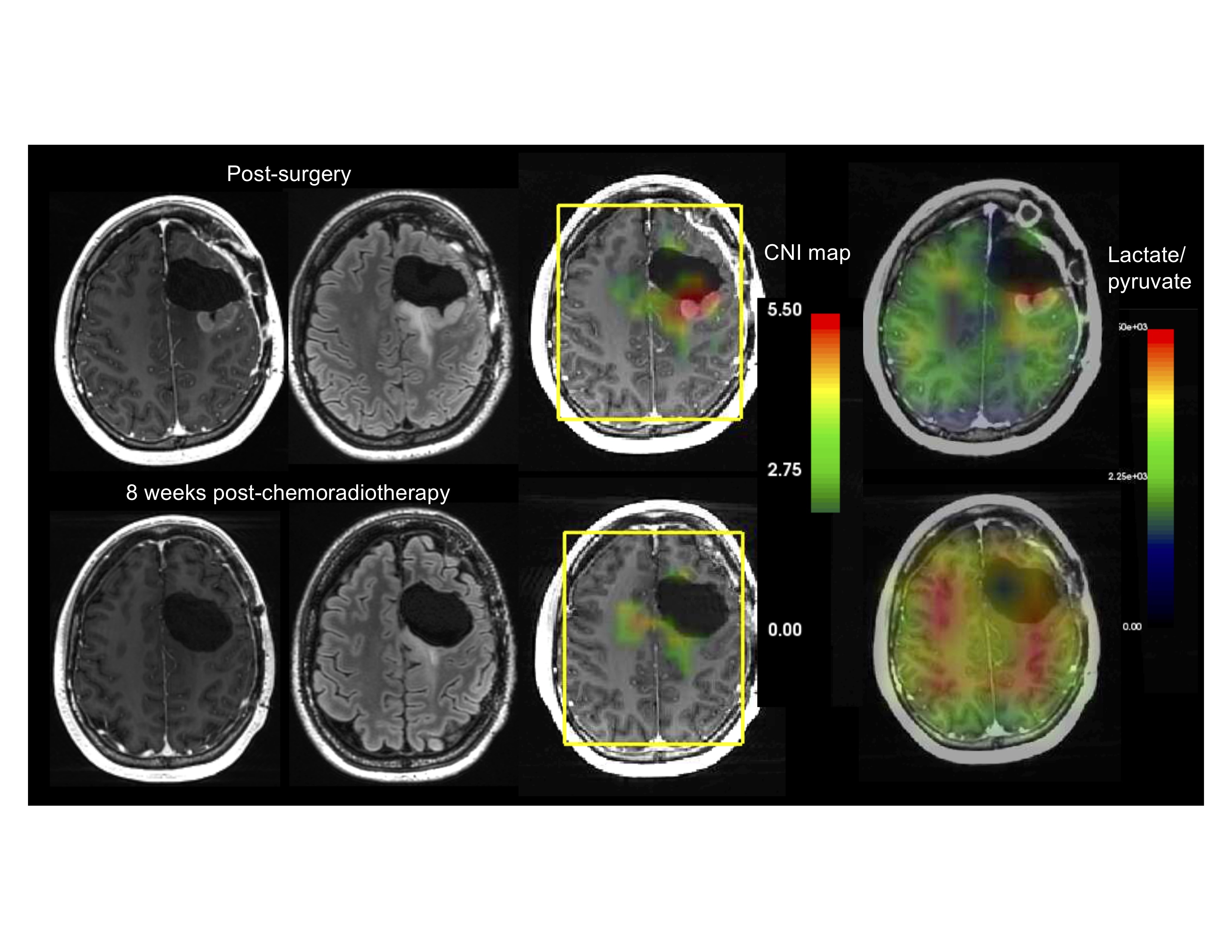
Marked response to chemoradiotherapy with resolution of contrast-enhancement within residual tumor and reduction in volume of FLAIR signal abnormality. Corresponding reductions in Choline to NAA index (CNI) and pyruvate-to-lactate conversion in 1H and 13C MRSI, respectively.
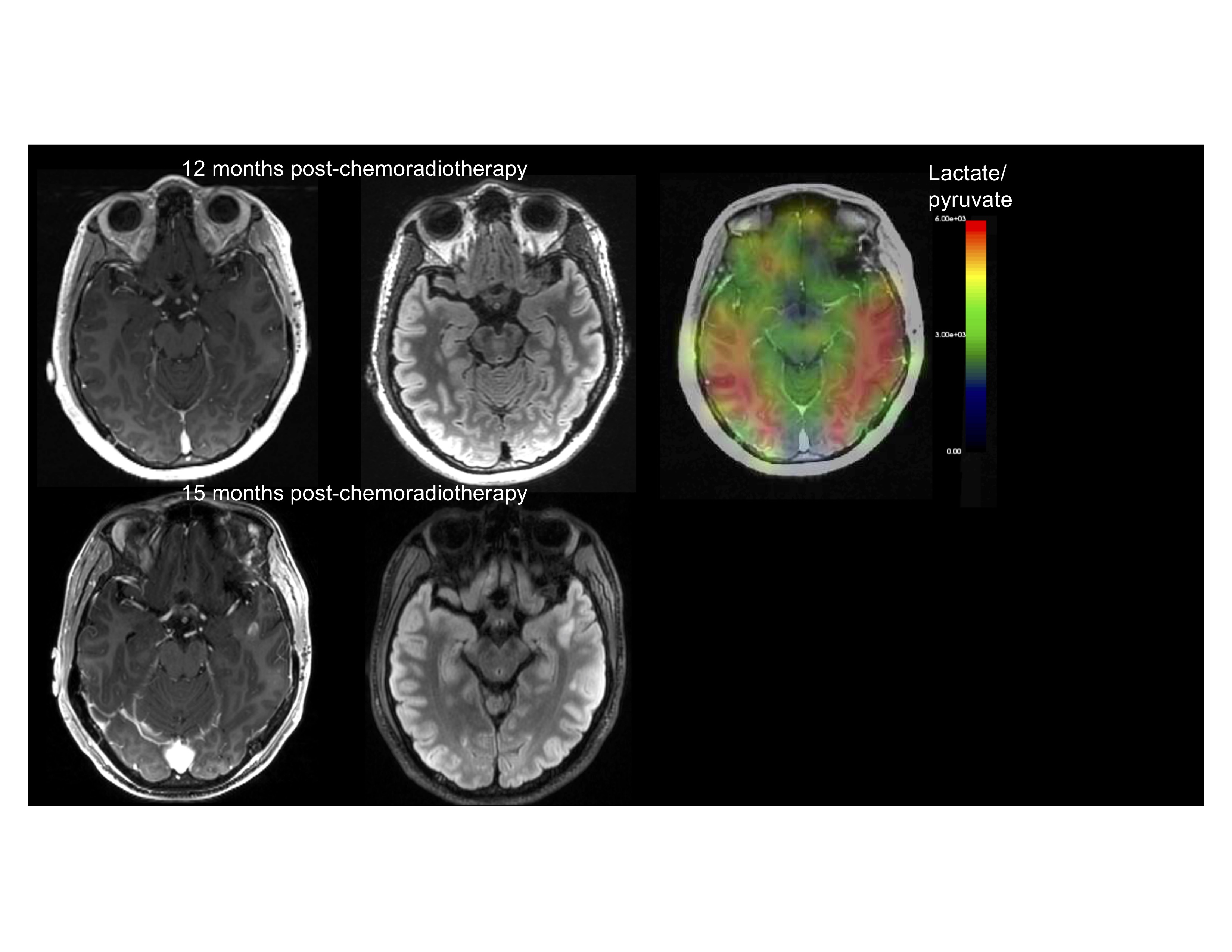
Increased pyruvate-to-lactate conversion in the left anterior temporal lobe in a region that subsequently develops contrast-enhancing recurrent glioma.
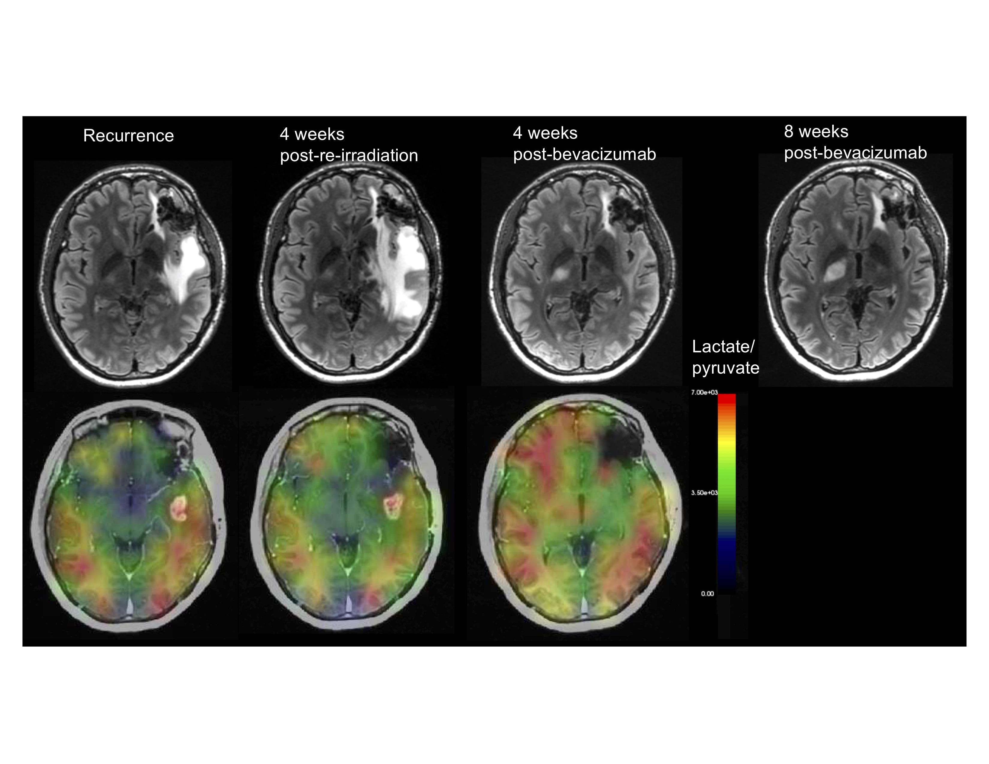
Increasing asymmetry of pyruvate-to-lactate conversion in the right internal capsule at a site of progressive recurrent glioma.
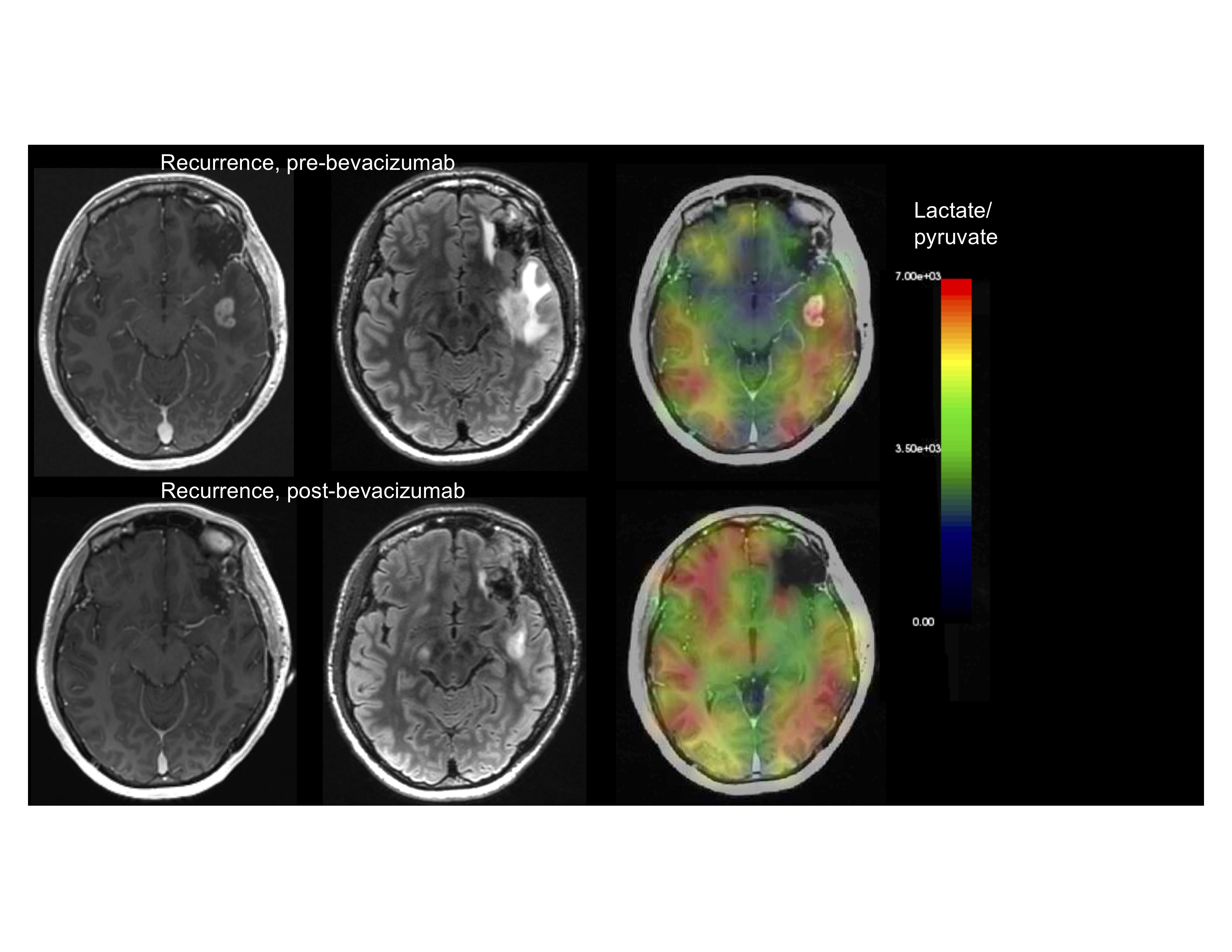
Decreasing contrast enhancement, FLAIR signal abnormality, and pyruvate-to-lactate conversion in a left temporal recurrent lesion following anti-angiogenic therapy (bevacizumab).