2863
Clinical and Radiological Characteristics of Histone H3 K27M Mutations in Midline Gliomas1Huaxi MR center(HMRRC), West China Hospital of Sichuan University, chengdu, China, 2Radiology, West China Hospital of Sichuan University, chengdu, China
Synopsis
The aim of the present study is to report the clinical and imaging characteristics of midline gliomas with H3 K27M mutations and to compare them with those gliomas without histone H3 K27M mutation in different age groups. There were 60 midline gliomas with H3 K27M mutation and 62 glioma patients without mutation included in this study. Mutant tumors were more likely diagnosed at an early age and located at infratentorial brain, with less contrast enhancement or edema.
Introduction
Somatic mutation in genes encoding the histone H3 always occurs in children and young adults with midline gliomas, such as glioma located in the brain stem and the spinal cord. H3 K27M mutations was used as a molecular feature to define the distinct entity, “Diffuse midline glioma, H3 K27M-mutant (grade IV)”, in the 2016 World Health Organization Classification of Tumors1. The characteristics of gliomas with H3 K27M mutations have been described in some researches2-3. Though, H3 K27M-mutant midline gliomas have no yet been compared with patients without histone H3 K27M mutation in detail.
Methods
We retrospectively enrolled patients with gliomas with or without H3 K27M mutations and reviewed their clinical and radiological characteristic. This study included 122 patients with midline gliomas, including 60 patients with H3 K27M mutation and 62 patients without H3 K27M mutation. The H3F3A/HIST1H3B mutation status was assessed in all patients, who underwent MR scanning.Results
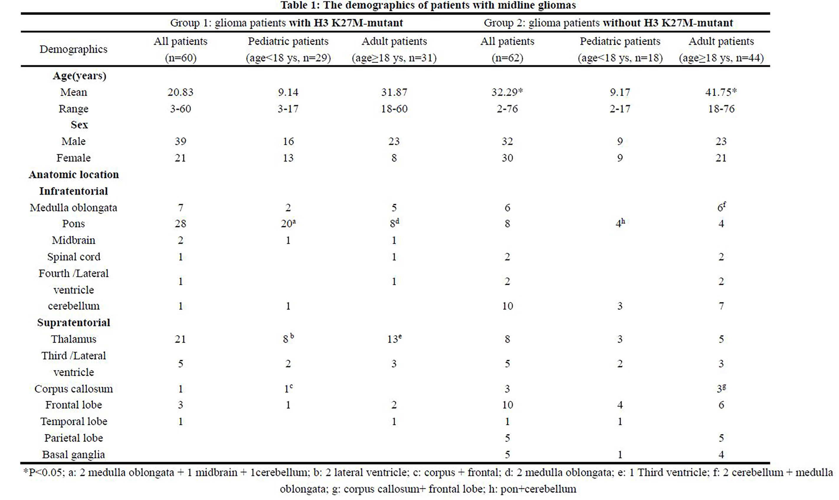
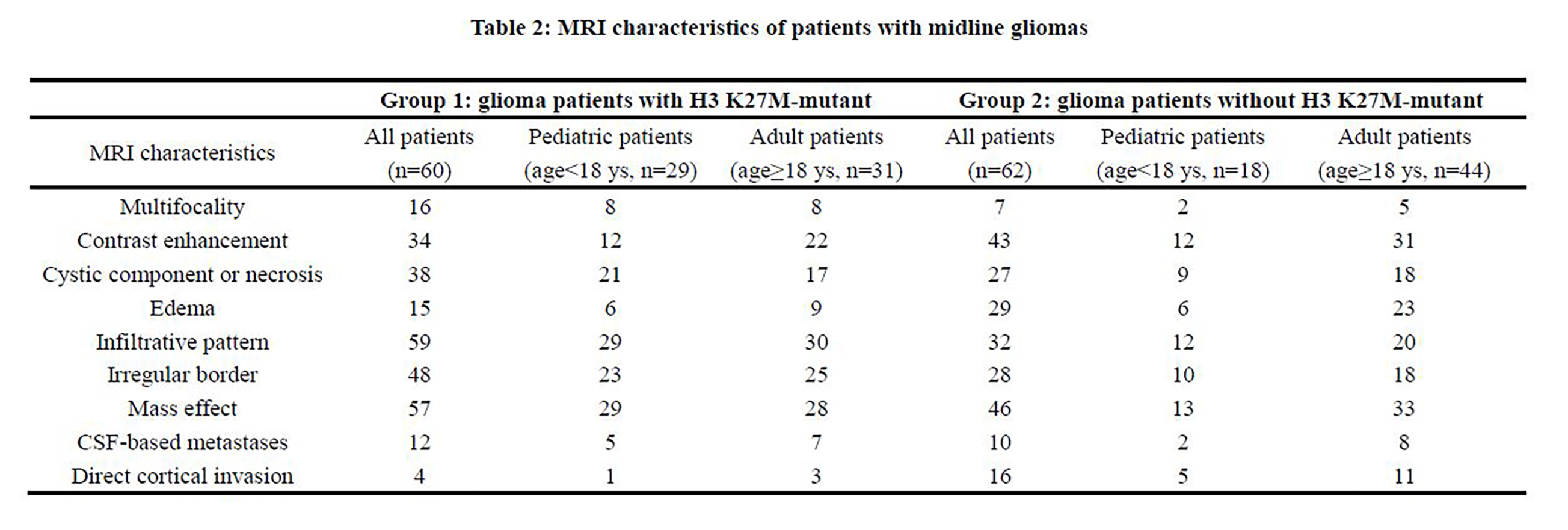
Their clinical characteristics are summarized in Tables 1. The median age at diagnosis in H3 K27M-mutant gliomas (mutant group) was 18 years (in pediatric: 9 y, range 3-17 y; in adult: 28 y, range 18-60 y). In group without mutation, patient age at time of diagnosis ranged from 2 to 76 years old with a median age of 34 years (in pediatric: 9 y, range 2-17 y; in adult: 38 y, range 18-76 y). There was significant difference between two groups. Male patients accounted for a greater proportion in both groups. Of the tumor’s location, in mutant group, all tumors located in medial structures of brain, 20 tumors in pediatric group were typically located in pons, and then followed by thalamus (n=8), medulla oblongata and ventricular system (co-occurred in the pons), cerebellum and frontal lobe. Though, in the adult group, most tumors are located in the thalamus (n=13), followed by pons (n=8), medulla oblongata (n=5, 2 co-occurred in the pons), ventricular system (n=4, 1 co-occurred in the thalamus), frontal and temporal lobe and spinal cord. And in group without mutation, tumors were located or next to the midline structure of brain, only 7 infratentorial tumors belong to pediatric patients, 4 tumors occurred in pons and 3 in cerebellum. And in supratentorial brain of pediatric patients, the frontal lobe was the most popular location (n=10), then followed by thalamus (n=8), third /lateral ventricle parietal lobe and basal ganglia (n=5, respectively), corpus callosum (co-occurred in the frontal lobe) and temporal lobe. In adult group, 21 tumors were located in infratentorial area, which were located in the cerebellum (n=7), medulla oblongata (n=6, co-occurred in the cerebellum), pons (n=4), ventricular system (n=2) and spinal cord (n=2), separately. Most tumors are supratentorial tumors, which are dispersed in the frontal (n=6), parietal (n=5, thalamus (n=5), basal ganglia (n=4), ventricular system (n=3), and corpus callosum. As for the radiographic features, in mutant group, most tumors were expansile masses with irregular border or necrosis, but little surrounding edema. Most tumors in adult patients demonstrated contrast enhancement (84%), only in 48% pediatric patients. In the group without mutation, most tumors demonstrated contrast enhancement and have mass effect, surrounded by edema. Besides, cystic component or necrosis and irregular border were common radiography characteristics.Discussion and Conclusion
H3 K27M-mutant gliomas were usually diagnosed at an earlier age than gliomas without mutation, a majority of tumors in both groups had a midline location. Pons and thalamus were the most common location in pediatric and adult patients in mutant group, respectively, though cerebellum and frontal lobe in group without mutation. Previous study has reported that the specific location on the midline seems to vary with age, mutant tumors are frequently located in pons in children, and the thalamus and the spine in adults2. Tumors have the different imaging appearances in two groups, mutant tumors always had mass effect, with less edema and contrast enhancement. But in tumors without mutation, most tumors demonstrated contrast enhancement, with large area of edema and cystic component or necrosis. There also had some similar imaging appearances in two groups, such as infiltrative pattern and mass effect. The diagnosis age, frequently locations and less contrast enhancement may consider as the characteristics to distinguish H3 K27M -mutant tumor in midline glioma.Acknowledgements
No acknowledgement found.References
1. Louis D.N., Perry A., Reifenberger G., von Deimling A., Figarella-Branger D., Cavenee W.K., Ohgaki H., Wiestler O.D., Kleihues P., Ellison D.W., 2016. The 2016 World Health Organization Classification of Tumors of the Central Nervous System: a summary. Acta neuropathologica 131, 803-820.
2. Solomon D.A., Wood M.D., Tihan T., Bollen A.W., Gupta N., Phillips J.J., Perry A., 2016. Diffuse Midline Gliomas with Histone H3-K27M Mutation: A Series of 47 Cases Assessing the Spectrum of Morphologic Variation and Associated Genetic Alterations. Brain pathology (Zurich, Switzerland) 26, 569-580.
3. Meyronet D., Esteban-Mader M., Bonnet C., Joly M.O., Uro-Coste E., Amiel-Benouaich A., Forest F., Rousselot-Denis C., Burel-Vandenbos F., Bourg V., Guyotat J., Fenouil T., Jouvet A., Honnorat J., Ducray F., 2017. Characteristics of H3 K27M-mutant gliomas in adults. Neuro-oncology 19, 1127-1134.
Figures