2861
The Initial Tumor Microenvironment in Cerebral Patient-Derived Glioma Xenografts Affects Their Phenotypical Presentation1Neurology, Henry Ford Health System, Detroit, MI, United States, 2Neurosurgery, Henry Ford Health System, Detroit, MI, United States, 3Radiology, Henry Ford Health System, Detroit, MI, United States, 4Radiation Oncology, Henry Ford Health System, Detroit, MI, United States, 5Public Health Sciences, Henry Ford Health System, Detroit, MI, United States
Synopsis
Patient-derived xenografts (PDXs) of human gliomas in murine models are unmatched in representing the molecular heterogeneity of the disease, but typically do not present with MRI contrast, and thus are not phenotypical. This may pose a limit to the assessment of trial therapies. PDX preparations were tweaked by co-injecting Matrigel with neurospheres. In one of the two cell lines studied, Matrigel promoted the formation of brain tumors whose genetic composition was that of the original human GBMs and whose radiologic appearance on MRI was similar to that seen in humans. In the second cell line, no BBB breakdown occurred.
Introduction:
In murine models, orthotopic patient derived xenografts (PDXs) generated from neurosphere cultures of human glioblastomas (GBMs) are unmatched in representing the molecular heterogeneity of the disease1. While these models have many advantages over traditional ones2, we and others3 have consistently observed that intraxial tumors with a blood-brain barrier (BBB) breakdown visible on MRI are extremely rare. Rather, typical phenotypes are infiltrative. This absence of phenotypic recapitulation of human tumors may lie in the interaction between tumor and stroma4. We hypothesized that manipulation of the tumor stromal composition would produce a consistent BBB breakdown in orthotopic murine models of GBM.Methods:
34 athymic nude rats were implanted intracerebrally with one of two neurosphere lines (HF2303 or HF3016) derived from two newly diagnosed GBM patients with identical time to progression (88 days) in which MRI studies showing typical ring-enhancing lesions.The transcriptional subclass (HF2303 is mesenchymal and HF3016 is proneural), and driver mutations in the original tumors are reproduced in the PDX. When the lesion exceeded 4 to 5mm in diameter, MRI studies determined whether BBB breakdown occurred. A board-certified neuroradiologist blinded to the conditions of the study reviewed all cases and graded each for radiological evidence of BBB breakdown on a scale of 0 to 4, with 0 being no evidence, and 4 being florid enhancement.
Tumor Preparation: In roughly equal numbers (13,11, and 10, respectively), PDX cells (3x10⁵cells in 10μl total solution) were co-injected with a preparation containing either 5μl Matrigel with growth factors (M+), 5μl Matrigel no growth factors (M-), or PBS as the control (M0).
MRI Studies: All studies used a Varian/Magnex(Santa Clara, CA), 7 Tesla, 20 cm bore magnet with a Bruker console running Paravision 6.0 software. Gradient maximum strengths and rise times were 250 mT/m and 120 ms. All MRI image sets were acquired with a 32x32 mm2FOV.Transmit and receive coils were Bruker Quadrature Birdcage and 4-channel phased-array surface coils. T1-weighted images (T1WIs) were acquired pre- and post-CA (Magnevist, 0.25 mmol/kg) with the following parameters: matrix: 256x192, 27 slices, 0.5 mm thickness, no gap, number of echoes (NE) = 1, number of averages (NA) = 4, TE/TR = 16/800 ms. To generate T2maps (T2Ms), a high-resolution spin-echo image was acquired pre-CA, with the following parameters: FA = 90°, 180°, matrix 256x192, 27 slices, 0.4 mm thickness, 0.1 mm gap, NE = 4, NA = 2, TE/TR = (20, 40, 60, 80)/3000 ms).
Results:
In 34 animals there were 5 extra-axial masses and 32 intra-axial cerebral tumors. All 5 extra-axial masses enhanced floridly. The 2 animals with only extra-axial masses were removed from further consideration.
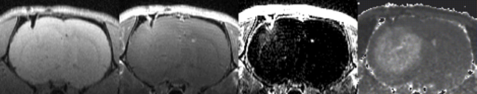
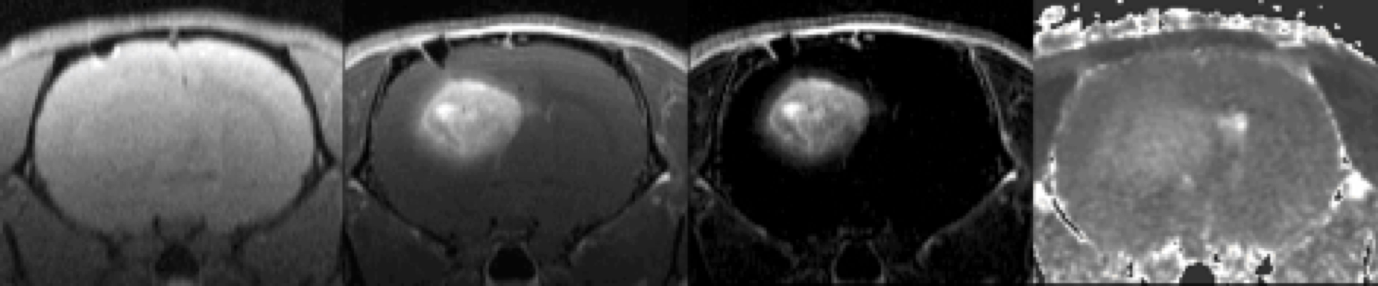
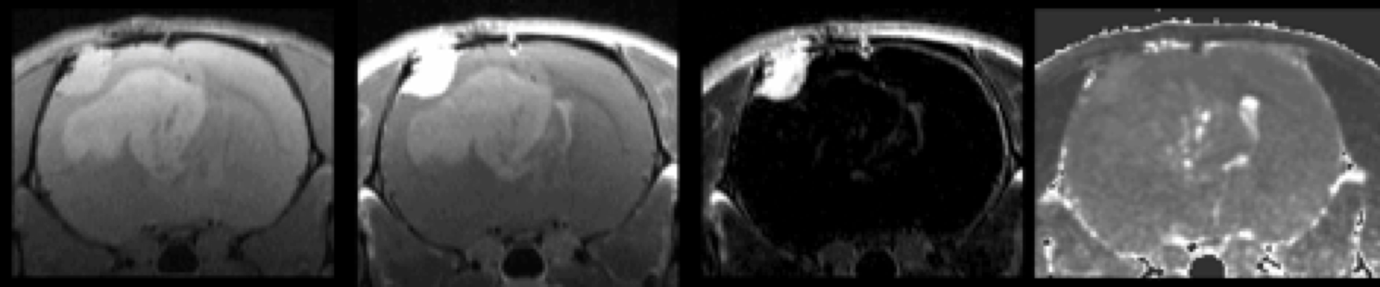
Visually, the two cell lines demonstrated quite different responses to Matrigel, with no subjective difference between M+ and M- in either cell line. Typical control HF3016 cell lines (M0) showed little or no T1WI post-Gd enhancement (Fig 1), and typical M+ and M- cell lines showed florid enhancement (Fig 2). Although T1WI HF2303 PDXs were easily visible both pre- and post-Gd (Fig 3), they showed little or no post-Gd enhancement under any condition (M0, M+, or M-).
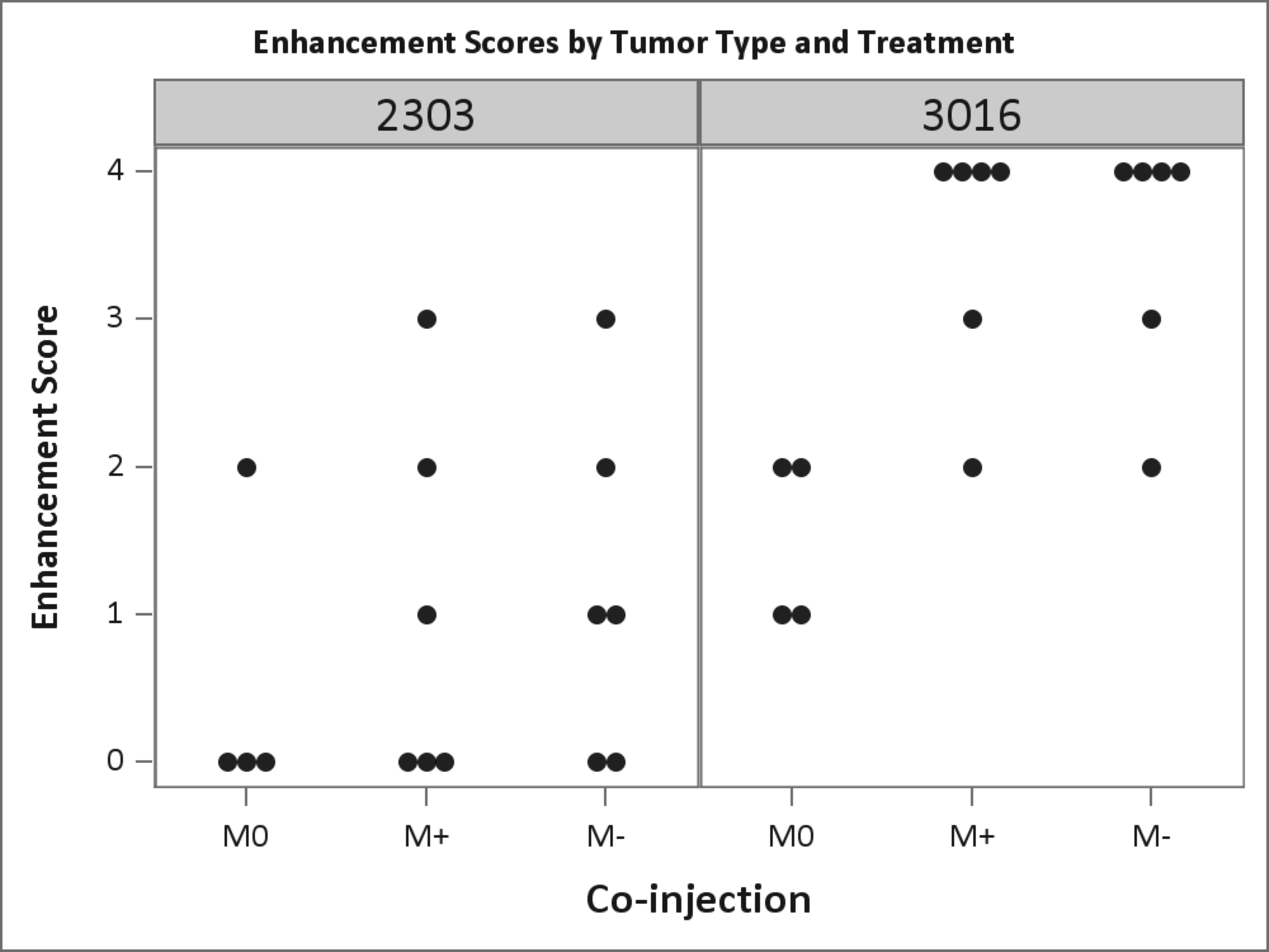
Statistical analysis supported these impressions.A dot plot (Fig 4) was used to visually summarize the data. A two-way ordinal logistic regression (OLR) model was fit with factors treatment (three levels: M0, M+ and M- ) and cell line (two levels HF2303 and HF3016). The test for interaction between cell line and M+ factor was not significant (p=0.451). There was also no evidence of a growth factor effect (p=0.725 for cell line HF2303 and p=1.0 for cell line HF3016). The Matrigel treated groups with and without growth factor were then combined. After this, the test for interaction between cell line and Matrigel was still not significant (p=0.195). However, when the Matrigel effect was tested using a Wilcoxon Rank Sum test separately within each cell line, the test was significant for HF3016 (N=16, p=0.004), but not for HF2303 (N=16, p=0.328).
Discussion/Conclusions:
As a practical matter for preclinical studies, a reliable BBB breakdown in PDX models promises a significant advance in the assessment of trial therapies because it produces an experimental condition much closer to the human pathology of interest. However, as demonstrated herein, the effect of stromal enhancement is cell-line dependent, and the biological correlates that accompany BBB breakdown still need to be described. An expansion of this investigation to other cell lines is underway, as is a molecular characterization of the cellular responses that accompany BBB breakdown after stromal enhancement.Acknowledgements
Funding was provided by Henry Ford Cancer Institute's Game on Cancer philanthropic funds.References
1.Vescovi, A.L., Galli, R. & Reynolds, B.A. Brain tumour stem cells. Nat Rev Cancer6, 425-436 (2006).
2. deCarvalho, A.C., et al.Gliosarcoma stem cells undergo glial and mesenchymal differentiation in vivo. Stem Cells28, 181-190 (2010).
3. Huszthy, P.C., et al.In vivo models of primary brain tumors: pitfalls and perspectives. Neuro Oncol14, 979-993 (2012).
4. Hidalgo, M., et al.Patient-derived xenograft models: an emerging platform for translational cancer research. Cancer Discov4, 998-1013 (2014).
Figures