2845
Structural changes in auditory and language-processing cortices and thalamus are predictors of word recognition ability after cochlear implantation1Neurology, University of Souther California, Los Angeles, CA, United States, 2Otolaryngology, Asan Medical Center, Seoul, Korea, Republic of
Synopsis
Introduction
Hearing loss is a common health problem, especially in the elderly. In adults with severe and profound hearing loss who do not benefit from hearing aids, cochlear implant (CI) is the only effective way to restore hearing [1]. However, speech perception ability after the surgery varies [2]. It is very important to build a computerized aiding system to assist clinicians in predicting postCI outcomes. Recent studies observed that long-term hearing loss in PD patients yielded structural changes in the cortical regions where auditory and language functions are processed [3]. These findings support the potential capacity of MRI morphological features in the prediction. Due to the complexity of multifactors (clinical variables and imaging markers) influencing the surgical outcome, nonlinear fitting algorithms may be suitable for this task. The purpose of our study is to predict word recognition scores (WRSs) as postCI outcome using a random forest regression (RFR) approach. Our study takes advantage of using the imaging features of structural MRI as well as conventional clinical parameters.Methods
The overall workflow is shown in Fig 1. We studied 100 patients with bilateral postlingual deafness (PD) and subsequently CI with full insert. A control group consists of 37 patients who experienced unilateral sudden hearing loss (SHL) but no other neurological issues. We used the following clinical variables as the predictors of post CI word recognition score (WRS): 1) duration of deafness (DoD); 2) age at CI (ageCI); 3) onset age of hearing loss (Onset). Presurgical MR imaging was performed using a 3T MRI scanner with an 8-channel coil. After a series of image processing including bias correction, brain masking, spatial normalization, and tissue segmentation, we obtained individual GM probability maps (smoothed with a 5mm FWHM Gaussian kernel). We used this voxel-wise map as the imaging features for the prediction of surgical outcome. We predicted WRS as postCI outcomes using RFR, a nonlinear fitting and supervised learning approach. To assess the ability of brain imaging features and clinical features in prediction, we performed three separate prediction tests using: 1) clinical feature-set, 2) imaging feature-set, and 3) combination of both feature-sets. To best utilize the brain regional features, we applied a data-driven cluster-based approach where the different combinations of the significant clusters were tested, and the best set leading to the highest accuracy was selected.Results
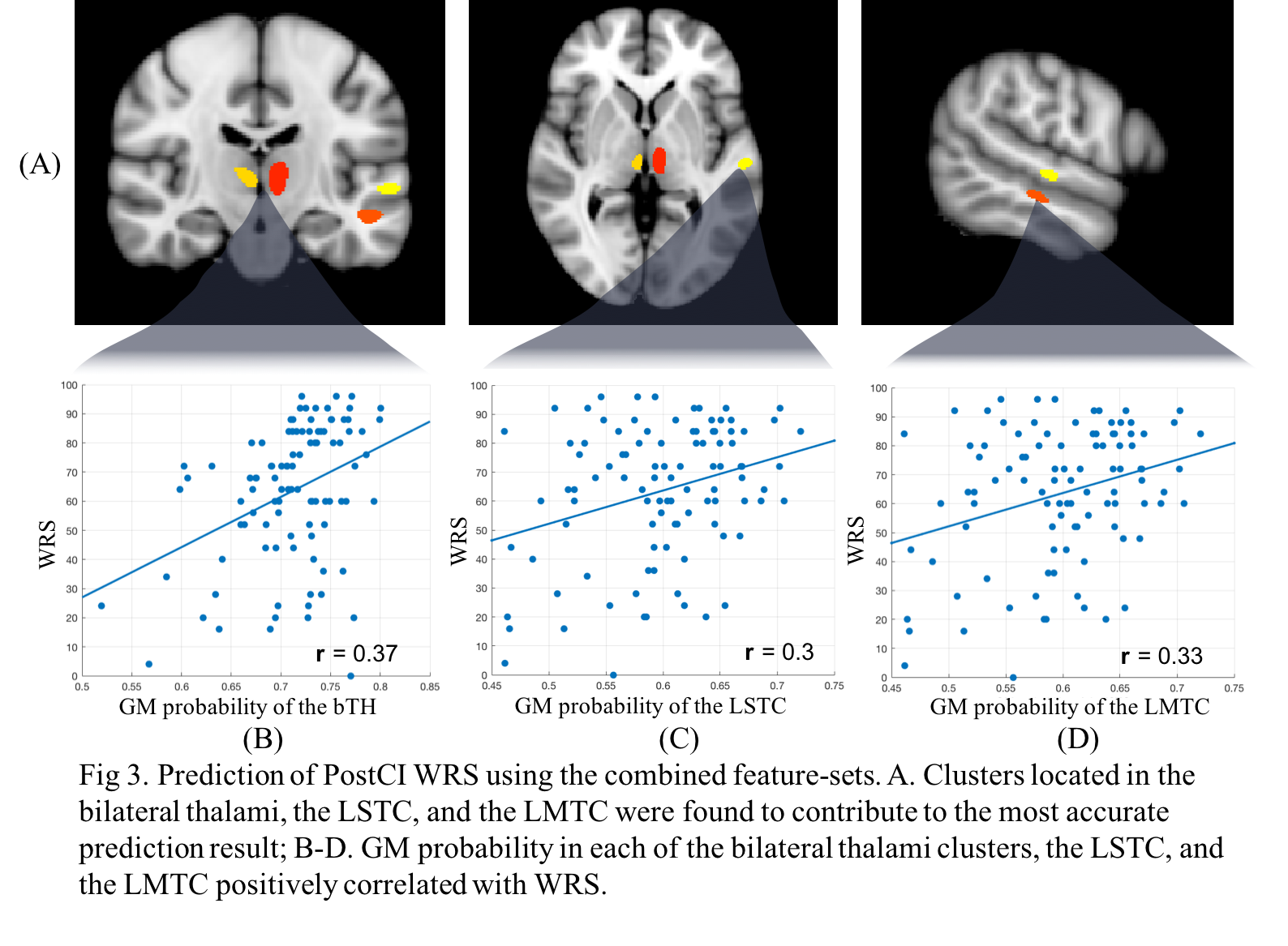
We found that the RFR prediction using the combined feature-sets of imaging and clinical features generated the most accurate prediction and outperformed either clinical or imaging feature-set alone (MAE: 13.8 vs.clinical: 16.60 vs imaging: 15.69; Fig 2). Across all the permutations, clusters contributing to the best prediction were located in the following three regions: bilateral thalami(bTH), left superior temporal (LSTC) and left middle temporal cortices (LMTC) (Fig 3A). Furthermore, we observed positive correlations between GM probabilities of these three regions, and WRS (r >=0.3; Fig 3B-D), i.e., the smaller their volumes were, the worse their surgical outcomes (= lower WRS) were. To evaluate the performance of RFR, we also predicted WRS using the linear SVMR method. We observed that RFR performed better than the linear SVMR, with a more marked difference when using the combined features (MAE difference: clinical: 4.5; imaging: 3.6; combined: 4.7; Table 1). In the RFR method, the computation of the feature importance showed that the thalamus contributed the most to the prediction (MAE increase when omitted: 7.27), followed by LMTC (3.98), DoD (3.49), ageCI (2.90) and Onset (1.00) (Fig 4).Discussion and Conclusion
We applied a RFR model to combined clinical and imaging features to predict postCI WRS, showing performance superior to a linear model. In this study, the GM probability in the two regions of bilateral TH and LMTC were the most important in prediction. These two regions biologically serve important roles in the language processing capacity which lacks in the PD subjects. Thalamic nuclei may control the networking between the fronto-opercular and temporo-cortical cortices, being responsible for the integration of lexico-syntactic and semantic information [4]. The LMTC, a critical node of the language network, is involved in the retrieval of lexical-syntactic information and comprehension of language [5]. Moreover, the correlation between the GM density of these two regions and the WRS suggests that progressive decreases in GM volume in patients with long-term post-lingual deafness may result in lack of recovery in language function after CI.Acknowledgements
No acknowledgement found.References
1. Cunningham LL, Tucci DL. Hearing loss in adults. New England Journal of Medicine. 2017;377(25):2465-73.
2. Green KM, Bhatt YM, Mawman DJ, et al.Predictors of audiological outcome following cochlear implantation in adults. Cochlear implants international. 2007 Oct;8(1):1-1.
3. Lazard, DS, Vincent, C, Venail, F, et al. Pre-, per-and postoperative factors affecting performance of postlinguistically deaf adults using cochlear implants: a new conceptual model over time. PloS one. 2012;7(11), e48739.
4. Klostermann F. Functional roles of the thalamus for language capacities. Frontiers in systems neuroscience. 2013;7:32.
5. Acheson DJ, Hagoort P. Stimulating the brain's language network: syntactic ambiguity resolution after TMS to the inferior frontal gyrus and middle temporal gyrus. Journal of Cognitive Neuroscience. 2013 Oct;25(10):1664-77.
Figures