2841
Predicting response to motor therapy in chronic stroke patients based on clinical and connectivity measurements using Machine Learning1Department of Radiology, Weill Cornell Medical College, New York City, NY, United States, 2Moss Rehabilitation Research Institute, Elkins Park, Elkins Park, PA, United States, 3Edith Cowan University, Joondalup, Australia, 4Burke Neurological Institute, White Plains, NY, United States, 5Iowa Neuroimaging and Noninvasive Brain Stimulation Laboratory, Departments of Pediatrics, Neurology & Psychiatry, University of Iowa Hospitals and Clinics, 200 Hawkins Drive, Iowa City, IA, United States, 6School of Electrical and Computer Engineering, and Meinig School of Biomedical Engineering, Cornell University, Ithaca, NY, United States, 7USC Neurorestoration Center, Los Angeles, CA; and Rancho Los Amigos National Rehabilitation Center, Downey, CA, United States, 8Brain and Mind Research Institute, Weill Cornell Medicine, New York, NY, United States
Synopsis
Statistical methods, including machine learning, are a highly promising avenue with which to improve prediction accuracy in clinical practice. The main objective of this study was to use machine learning methods to predict a chronic stroke individual’s motor function after 6 weeks of intervention from demographic, neurophysiological and imaging measurements. Our main finding was that Elastic-net outperformed Support Vector Machine, Artificial Neural Network, Random Forest, and Classification and Regression Trees in predicting post-intervention Fugl-Meyer Assessment. The addition of structural dysconnectivity measurements to the demographic and neurophysiological data did not improve the performance of the methods.
Introduction
Accurate predictions of motor improvement resulting from intensive therapy in chronic stroke patients is a difficult task for clinicians but are key in prescribing appropriate therapeutic strategies.1,2 Statistical methods, including machine learning, are a highly promising avenue with which to improve prediction accuracy in clinical practice.3,4 The first main objective of this study was to use machine learning methods to predict a chronic stroke individual’s motor function after 6 weeks of intervention from demographic, neurophysiological and imaging measurements. The second main objective was to identify which measurements were most important in predicting chronic stroke patients’ impairment after 6 weeks of intervention.Methods
Data from one hundred and two patients (Female: 31%, age 61±11 years) who suffered first ischemic stroke 3-12 months prior were included in this study. After enrollment, patients underwent 6 weeks of intensive motor and transcranial magnetic stimulation therapy. Age, gender, handedness, time since stroke, pre-intervention Fugl-Meyer Assessment, stroke lateralization, difference in motor threshold between the unaffected and affected hemispheres, absence of motor evoked potential in the affected hemisphere and various imaging metrics (regional and pair-wise structural disconnectivity) were used as predictors of post-intervention Fugl-Meyer Assessment. Five machine learning methods (Elastic-Net, Support Vector Machine, Artificial Neural Networks, Decision Trees, and Random Forest) were used to predict post-intervention Fugl-Meyer Assessment based on either demographic and neurophysiological measurements alone or in combination with the imaging metrics. Cross-validated R-squared and root of mean squared error were used to assess the prediction accuracy and compare the performance of methods.Results
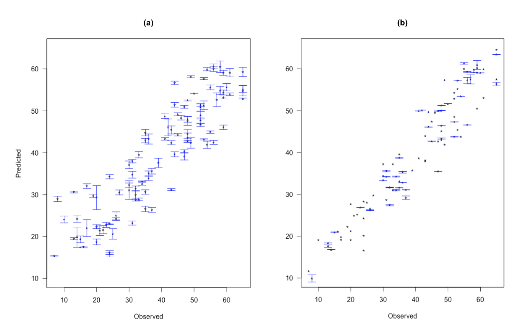
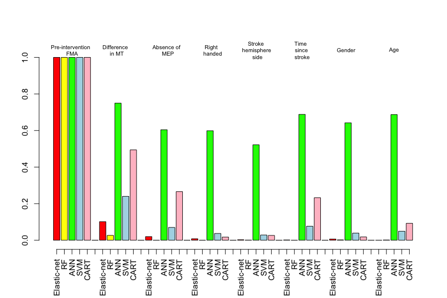
Elastic-net performed significantly better than the other methods for the model containing pre-intervention Fugl-Meyer Assessment, demographic and neurophysiological measurements as predictors of post-intervention Fugl-Meyer Assessment; p < 0.05). CART and EN showed respectively the lowest and the highest R2 results with the clinical variables. It is important to note that the standard deviation in the individual predictions calculated using the CART model were much larger when compared to the EN model predictions (See Figure 1). Pre-FMA and difference in motor threshold were commonly found as the strongest two predictors in the clinical model (See Figure 2). The difference in motor threshold had greater variable importance than the absence of motor evoked potential in the affected hemisphere for the five methods.Discussion
Our main finding was that Elastic-net out performed other methods in the regression analysis. The addition of both structural dysconnectivity measurements to the demographic and neurophysiological data did not improve the performance of the methods compared to the model including demographic and neurophysiological data only.Conclusion
The approach implemented here may enable clinicians to more accurately predict a chronic stroke patient’s individual response to intervention. The predictive models used in this study could assist clinicians in making treatment decisions and improve the accuracy of prognosis in chronic stroke patients, a notoriously difficult task. Both functional and structural connectivity metrics that are measured at acute and chronic stage can be used in a future study to characterize better the brain injury.Acknowledgements
No acknowledgement found.References
Kim, B.; Winstein, C. Can Neurological Biomarkers of Brain Impairment Be Used to Predict Poststroke Motor Recovery? A Systematic Review. Neurorehabilitation and Neural Repair 2017, 31, 3–24.
Nijland, R. H. M.; Wegen, E. E. H. van; Harmeling-van der Wel, B. C.; Kwakkel, G. Accuracy of Physical Therapists’ Early Predictions of Upper-Limb Function in Hospital Stroke Units: The EPOS Study. Physical Therapy 2013, 93, 460–469.
Cohen, J. R.; Asarnow, R. F.; Sabb, F. W.; Bilder, R. M.; Bookheimer, S. Y.; Knowlton, B. J.; Poldrack, R. A. Decoding Continuous Variables from Neuroimaging Data: Basic and Clinical Applications. Frontiers in Neuroscience 2011, 5, 1–12.
Wang, Y.; Fan, Y.; Bhatt, P.; Davatzikos, C. High-Dimensional Pattern Regression Using Machine Learning: From Medical Images to Continuous Clinical Variables. NeuroImage 2010, 50, 1519–1535.
Figures