2827
Quad-contrast imaging with deep learning-powered reconstruction: 2-minute neuro-evaluation1Electrical and Computer Engineering, Seoul National University, Seoul, Korea, Republic of, 2Biomedical Engineering, Hankuk University of Foreign Studies, Seoul, Korea, Republic of
Synopsis
A 2D multi-contrast sequence with deep learning-powered reconstruction is developed to generate four contrast images (PDw, T1w, T2w, and FLAIR) and two quantitative maps (T1 and T2) in 2 minutes of scan time. For the reconstruction, a new deep learning method that assures both data consistency and image fidelity is applied with the joint reconstruction of the quad-contrast k-space data.
Introduction
Despite recent advances in 3D high-speed high-resolution imaging, 2D imaging for T2w, FLAIR, T1w, and PDw are common practice for a routine neuro-evaluation in clinic. These 2D data are acquired with limited number of slices (~20) and thick slices (~5 mm), still requiring the total scan time of 10+ min. Hence, reducing this scan time has an important practical impact. Multi-contrast imaging that acquires multiple contrasts in a single scan is an attractive solution for reducing scan time. We recently proposed a novel 2D multi-contrast imaging sequence1, which produces the four contrasts and T1 and T2 maps at the scan time of 7 minutes. In this work, the scan time is further reduced to 2 min by incorporating a deep learning-powered reconstruction. To benefit from the shareable information among the four contrasts, undersampling schemes were devised for joint reconstruction of the multiple contrasts2.Methods
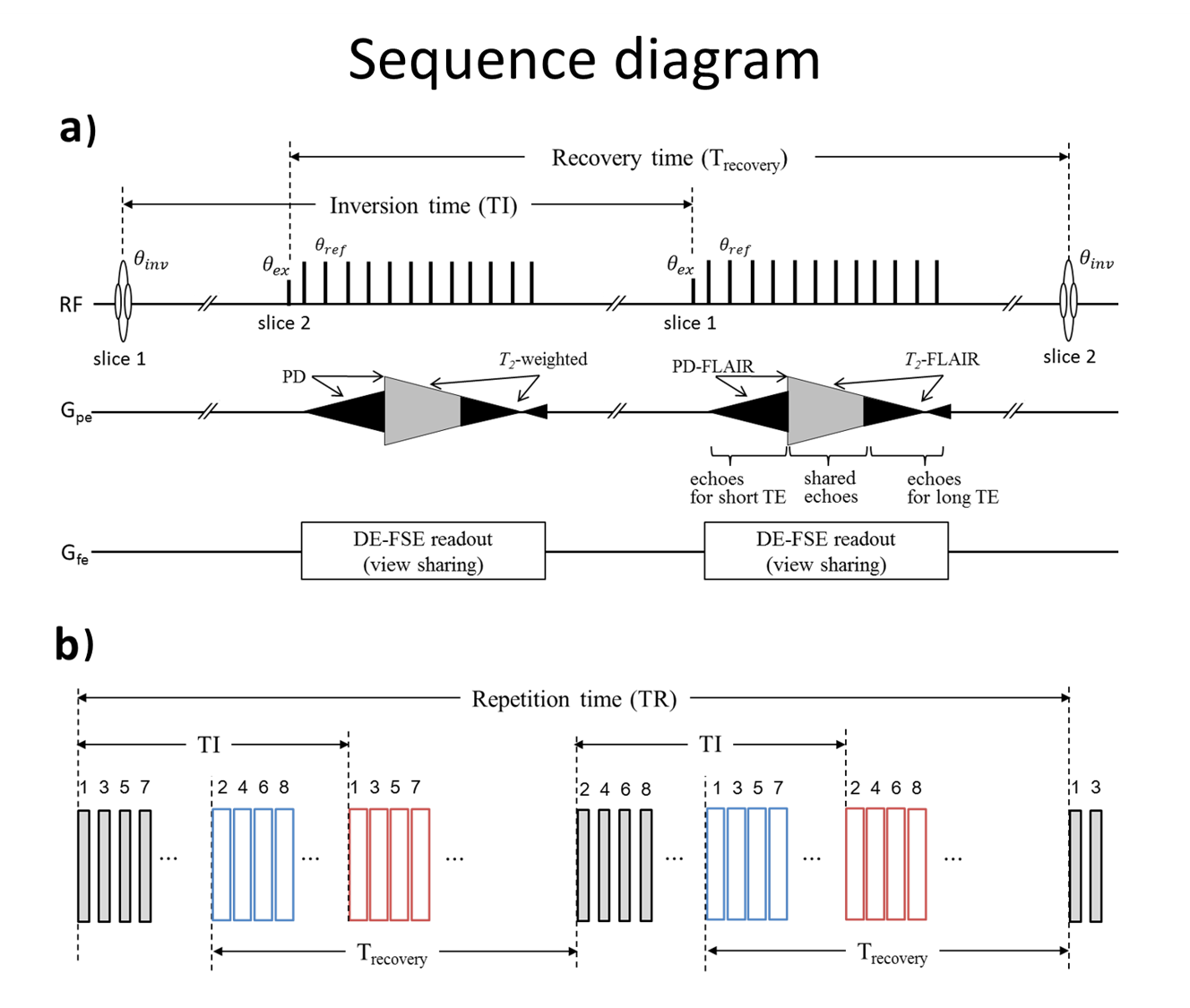
[Quad-contrast sequence] The 2D quad-contrast sequence (Figure 1) was employed to acquire four native contrasts, PDw, T2w, PD-FLAIR and T2-FLAIR. From these contrasts, T1w images, and T1 and T2 maps were generated. T1w images were calculated by the ratio of the PD-FLAIR and PDw. For the T1 map, the signal evolution of the PD and PD FLAIR images were formulated:
$$S_{PD}= ρ(1-e^{-T_{recovery}/T_1 })$$
$$S_{PD-FLAIR}= ρ(1-2e^{-TI/T_1}+e^{-T_{recovery}/T_1 } e^{-TI/T_1 })$$
Then, T1 maps were fitted using non-linear least squares fitting such that errors are minimized for both equations. The PD and T2w images were used to fit T2 maps. The fitting was performed using an extended phase graph dictionary with a T2 value range from 1 ms to 300 ms and 0.1 ms resolution.
[Data acquisition] Full k-space data were acquired using the quad-contrast
sequence. The scan parameters were FOV = 256 × 256 mm2, voxel size = 1 × 1 mm2,
slice thickness = 5 mm, number of slices = 20, TR = 12000 ms, TI = 2000 ms and
total scan time = 6.4 min. Ten healthy subjects were scanned at 3T.
The fully sampled data were uniformly
undersampled in the phase encoding direction by a factor of 4 with 32
autocalibration lines. To benefit from the shared information among the four
different contrasts, undersampling schemes were devised. For the PD and T2w contrasts,
k-space lines were undersampled uniformly starting from the second line, and for
the PD-FLAIR and T2-FLAIR contrasts, k-space lines were undersampled starting from
the fourth line.
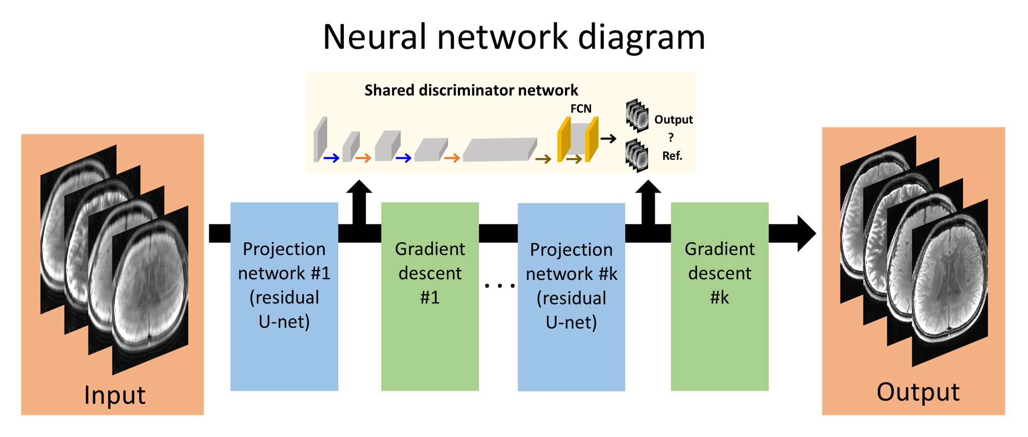
[Deep learning based reconstruction] A new deep learning-powered image
reconstruction that enforces both data consistency and image fidelity3
was modified to
jointly reconstruct the four contrasts by receiving all four undersampled
contrasts as input and generating four fully sampled contrasts as output
(Figure 2). Seven datasets out of the ten subject scans were used for network
training, one for validation, and two for test. To overcome the lack of data,
data augmentation was done by flipping the image (total training image patch:
7840).
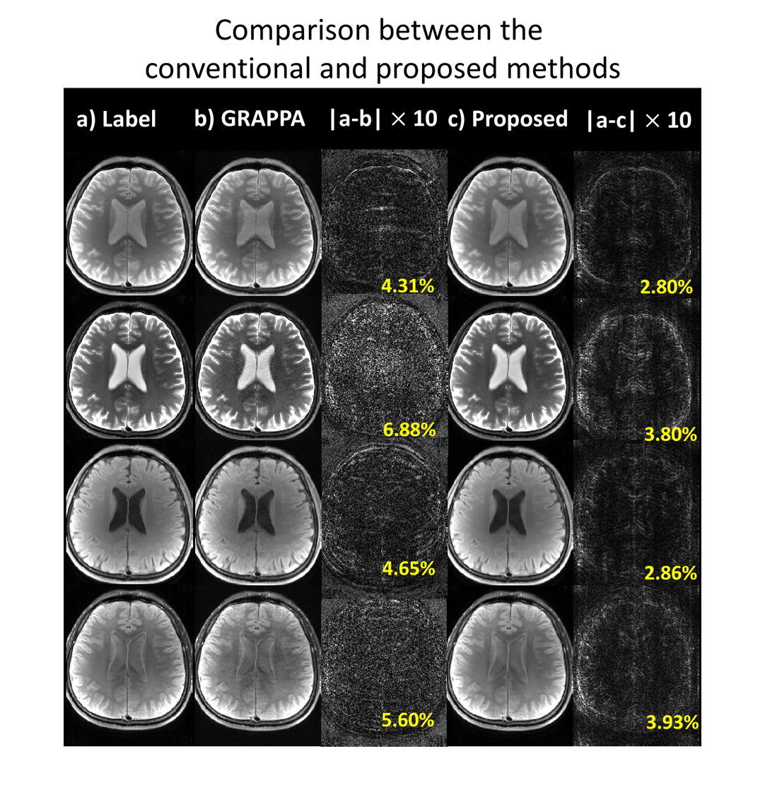
To examine the quality of the reconstructed images,
the results were compared with images reconstructed with GRAPPA4.
Results
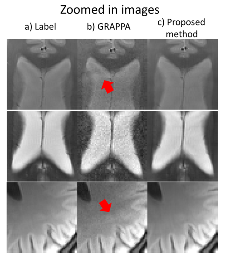
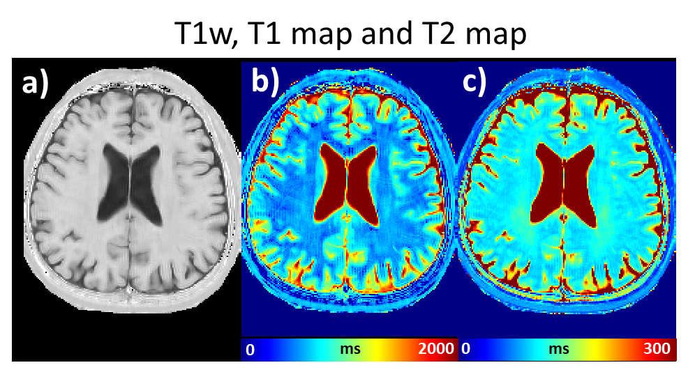
The four contrast images are shown in Figure 3 for the full-sampled data reconstruction (Figure 3a; label), GRAPPA reconstruction (Figure 3b), and proposed reconstruction (Figure 3c). The results show superior image quality and lower nRMSE in our method (Figure 3c) when compared to GRAPPA (Figure 3b). In the zoomed-in images (Figure 4), significant decrease in aliasing artifacts can be observed. Figure 5 shows the processed T1w image, T1 map and T2 map, revealing high image quality.Conclusion and Discussion
In this work, we proposed a quad-contrast sequence accelerated by a deep learning-powered reconstruction for 2 minute neuro-evaluation. The four contrasts, T2w, FLAIR, PD and T1w that are routinely utilized in clinic were generated along with T1 and T2 maps. Different from recent multi-contrast methods such as MRF5 or MAGiC6, our T2w, FLAIR and PD images are not processed images but natively acquired from the sequence. Hence, they can be directly applied in clinic as a routine neuro-evaluation. Because undersampling the full k-space is equivalent to choosing a few shots in the sequence, the implementation of this method is straightforward. Further improvement in reconstruction may be achieved by randomizing the undersampling pattern.Acknowledgements
This work was supported by Creative-Pioneering Researchers Program through SNU and Brain Korea 21 Plus Project in 2018References
1. Jung JH, Penta-contrast imaging: a Novel Pulse Sequence for Simultaneous Acquisition of Proton Density, T1, T2, T2* and FLAIR images. ISMRM 2017
2. Gong EH, Huang F, Ying K, Wu WC, Wang S, Yuan C. PROMISE: Parallel-Imaging and Compressed-Sensing Reconstruction of Multicontrast Imaging Using SharablE Information. Magnetic Resonance in Medicine 2015;73:523-+.
3. Lee DH, MRI acceleration using projected gradient descent with iterative shared-discriminator GAN. Submitted in ISMRM 2019
4. Griswold MA, Jakob PM, Heidemann RM, et al. Generalized autocalibrating partially parallel acquisitions (GRAPPA). Magn Reson Med 2002;47:1202-1210.
5. Ma D, Gulani V, Seiberlich N, et al. Magnetic resonance fingerprinting. Nature. 2013;495(7440):187-92.
6. L.N. Tanenbaum, et al. Synthetic MRI for Clinical Neuroimaging: Results of the Magnetic Resonance Image Compilation (MAGiC) Prospective, Multicenter, Multireader Trial. (2017) American Journal of Neuroradiology. 38 (6): 1103.
Figures