2815
GABA and GSH at 7T: to edit or not to edit?1Danish Research Centre for Magnetic Resonance, Centre for Functional and Diagnostic Imaging and Research, Copenhagen University Hospital Hvidovre, Hvidovre, Denmark, 2Center for Magnetic Resonance, Dept. Electrical Engineering, Technical University of Denmark, Lyngby, Denmark
Synopsis
With the number of 7T sites increasing, more brain-MRS studies will be conducted at 7T. Particularly GABA and GSH are of interest and benefit from the increased resolution. To date, studies have applied both conventional and edited MRS to measure these metabolites, however, there is no consensus on whether to use edited MRS. This study shows that it depends on brain region and potentially voxel size and number of acquisitions, whether it is preferable to use editing for GABA. There is less reason to argue that editing benefits measurement of GSH, but this study indicates a slight preference for editing.
Introduction
With the number of 7T sites rapidly increasing, more brain-MRS studies will be conducted at 7T. Particularly GABA and GSH are of interest, as these metabolites have small signals that are overlapped by larger signals, and benefit from the increased resolution at 7T. To date, studies have applied both conventional and edited MRS to measure GABA and GSH at 7T. However, there is no consensus on whether to use edited MRS for measurement of GABA and GSH at 7T. This study is a first attempt to compare conventional and edited MRS methods for measurement of GABA and GSH at 7T.Methods
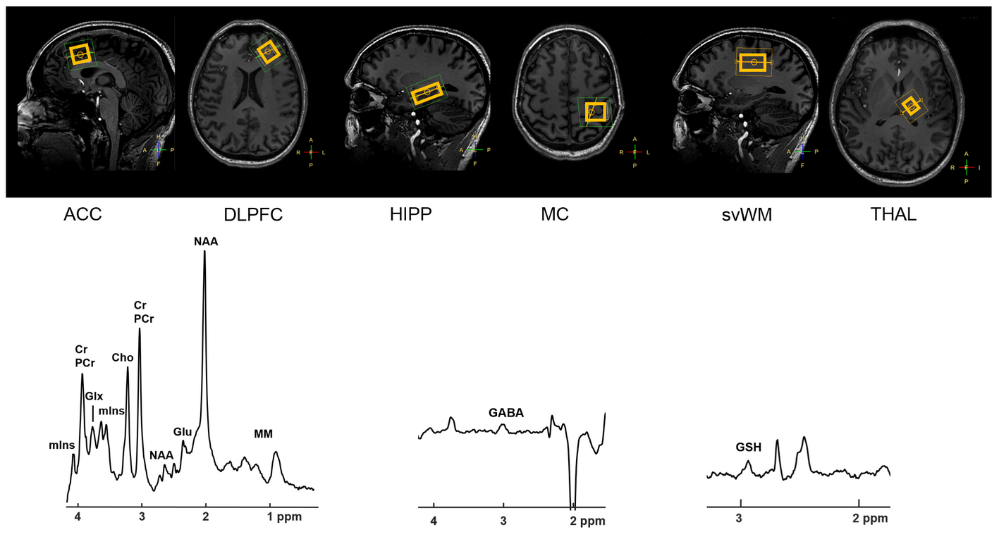
15 healthy volunteers (age 22.7±2.5, M/F=7/8) were scanned twice with four days between sessions. Conventional (sLASER1,2) and edited (Hadamard edited sLASER2,3) MRS were acquired in the anterior cingulate cortex (ACC), dorsolateral prefrontal cortex (DLPFC), hippocampus (HIPP), motor cortex (MC), supraventricular white matter (svWM) and thalamus (THAL) (Fig.1). Experiments were performed in accordance with local ethical guidelines.
MRS experiments (see Table 1 for sequence parameters and Fig. 1 for example spectra) were performed with a 7T MR scanner (Philips, Best, The Netherlands) in combination with a dual transmit coil and 32-channel receive coil (Nova Medical, Wilmington, MA, USA). Automated planning was used to ensure uniform voxel placement throughout the study.
sLASER spectra were fitted in LCModel4 using a basis set including 20 metabolites and a macromolecular baseline. Edited spectra were fitted using in-house developed software implemented in Matlab (The Mathworks Inc., Natick, MA, USA). Metabolite concentrations were corrected for partial volume effects5.
Statistical analyses were performed with SPSS 22 (IBM Corp., Armonk, NY, USA). Only concentration estimates with a CRLB of <20% were included in the analyses. Paired t-tests were used to test differences between conventional and edited concentration estimates in session 1, between conventional concentration estimates in session 1 and 2, and between edited concentration estimates in session 1 and 2.
Results
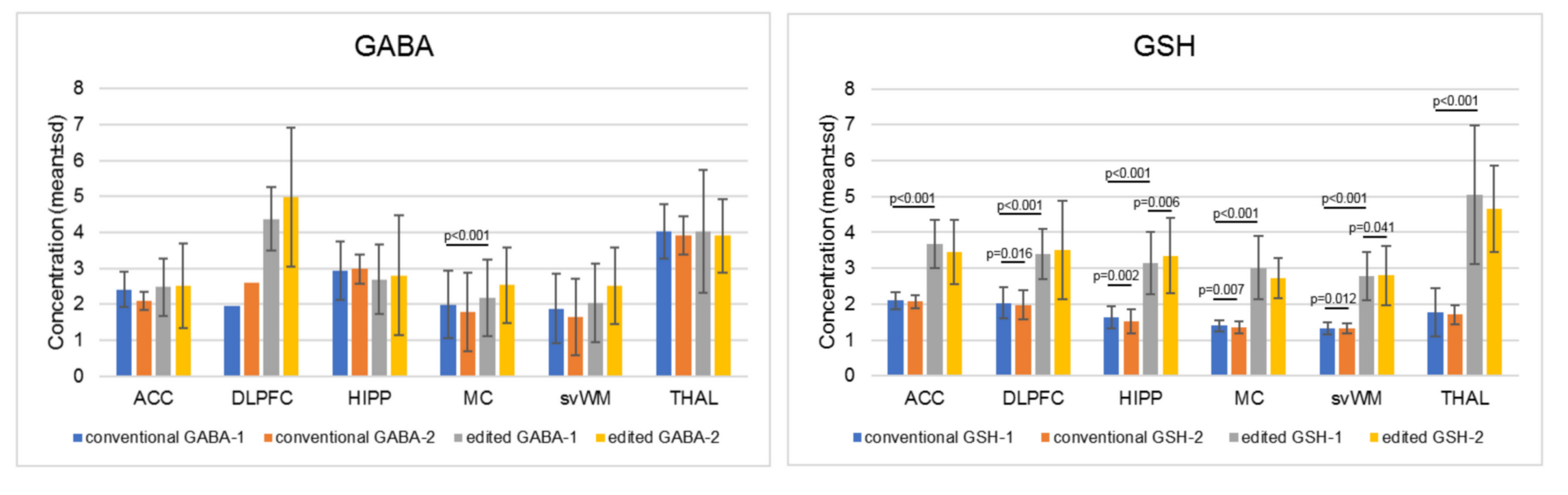
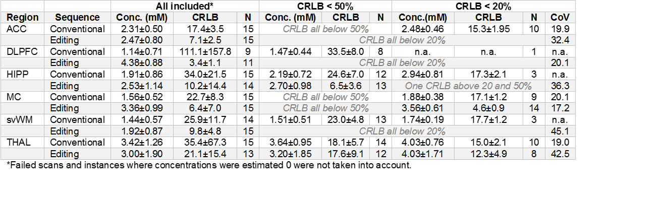
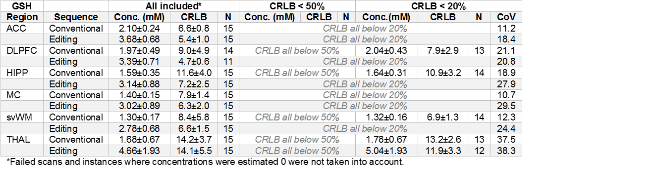
Tables 2 and 3 describe conventional and edited concentration and CRLB estimates and CoV’s for GABA and GSH. Fig. 2 describes concentration estimates for all measurements.
For GABA, conventional and edited concentration estimates significantly differed for MC (p<0.001) but not for other regions (DLPFC did not yield enough usable data). For GSH, conventional and edited concentration estimates significantly differed for all regions (p<0.001).
Between session 1 and 2, conventional GSH concentration estimates for DLPFC (p=0.016), HIPP (p=0.002), MC (p=0.007) and svWM (p=0.012) significantly differed; no significant differences were observed for ACC and THAL. No significant differences were observed for conventional GABA concentration estimates for ACC, MC and THAL (DLPFC, HIPP and svWM did not yield enough usable data).
Between session 1 and 2, edited GSH concentration estimates for HIPP (p=0.006) and svWM (p=0.041) significantly differed; no significant differences were observed for ACC, DLPFC, MC and THAL. No significant differences were observed for edited GABA concentration estimates for any region.
Discussion
GABA: Editing resulted in more included data based on CRLB for all regions except THAL, whereas CoV’s were higher for edited MRS. Particularly for DLPFC, HIPP and svWM, a large amount of data was excluded from the conventional MRS measurements based on CRLB<20%, whereas this was not the case for edited MRS. For svWM, this could be because GABA concentrations are low in white matter and may therefore be easier to measure with edited MRS. Conventional and edited MRS yielded similar results in all regions except MC, where edited concentration estimates were almost twice as high as compared to conventional concentration estimates. There were no differences between sessions for edited GABA, suggesting reasonable agreement across regions.
GSH: Editing consistently resulted in higher estimated concentrations. CoV’s were almost consistently higher for edited compared to conventional MRS, except for DLPFC and THAL where similar CoV’s were observed. Overall, editing did not result in more included data. Conventional and edited concentration estimates differed in all regions, suggesting low agreement between the two methods, possibly due to the fact that editing yields higher concentration estimates. Differences between sessions were observed in four regions for conventional MRS and in two regions for edited MRS.
These analyses are part of an ongoing study which is expected to finish medio 2019.
Conclusion
Whether it is preferable to use edited MRS for measurement of GABA at 7T depends on brain region and potentially voxel size and number of acquisitions. Particularly measurements in DLPFC, HIPP and svWM may benefit from editing. There is less reason to argue that editing benefits measurement of GSH at 7T, but the preliminary analyses from this study indicate a slight preference for edited MRS.Acknowledgements
This research is supported by the Danish Agency for Science, Technology and Innovation grant no. 0601-01370B, and The John and Birthe Meyer Foundation.References
1. Boer et al. (2011), NMR Biomed, 24(9):1038-1046. 2. Arteaga de Castro et al. (2013), NMR Biomed, 26(10):1213-1219. 3. Saleh et al. (2018), Magn Reson Med, 80(2):474-479. 4. Provencher (1993), Magn Reson Med, 30(6):672-679. 5. Gasparovic et al. (2009), J Neurotrauma, 26(10):1635-1643.Figures