2792
Quantitative Susceptibility Mapping shows differences in substantia nigra of individuals with 22q11.2 deletion syndrome and controls1Electrical Engineering, Pontificia Universidad Católica de Chile, Santiago, Chile, 2Biomedical Imaging Center, Santiago, Chile, 3Millennium Nucleus for Cardiovascular Magnetic Resonance, Santiago, Chile, 4Biomedical Imaging Center, Santiago, Chile, Santiago, Chile, 5Department of Radiology, Pontificia Universidad Católica de Chile, Santiago, Chile, 6Wellcome Centre for Human Neuroimaging, University College London, London, United Kingdom, 7Center of Genetics and Genomics, Santiago, Chile
Synopsis
Unlike individuals with Parkinson’s disease, patients with 22q11.2 deletion syndrome at risk of Parkinson show an increase in dopamine at striatal regions. Since iron levels are related to dopamine levels, we studied the difference of magnetic susceptibility between 17 patients with the deletion and 19 healthy individuals. Susceptibility measurements were obtained with QSM and then compared using a Mann Whitney U test. Results showed a significant difference in the substantia nigra, which indicates a possible cause for the increased levels of dopamine in 22q11.2 individuals at Parkinson’s risk.
Introduction
The 22q11.2 Deletion Syndrome (22q11.2DS) is a neurogenetic disorder resulting from a microdeletion of approximately 3 megabases on the long arm of chromosome 221. It results in a heterogeneous clinical presentation, being at higher risk of neuropsychiatric disorders, particularly psychosis1,2. Recently, the 22q11 deletion has also been found to confer a high risk for early-onset Parkinson’s disease. As such, it is likely that these patients have a dopaminergic abnormality at the core of the disorder. A previous study used PET images with the radioligand 11C-DTBZ that binds to a protein that transports cytosolic dopamine to study the dopaminergic system in these patients. They found an increase of the radioligand binding in patients at risk of Parkinson’s disease3. There is a pressing need to continue characterizing the dopaminergic system in 22q11DS patients.
Neuromelanin, a by-product of dopamine metabolism, also chelates iron. On the other hand, MRI-based quantitative susceptibility mapping (QSM) has shown to be sensitive to iron content4. As such, we here used QSM to examine the dopaminergic system in individuals with 22q11.2 DS.
Methods
A total of 36 individuals participated in the study. The comparison groups comprised 17 individuals with 22q11.2DS and 19 healthy controls. The images acquired were magnitude, high resolution T1w 3D images and phase and magnitude images obtained from a Gradient-recalled multi-echo (GRE) sequence with 5 echoes. First TE = 7.2ms, ΔTE = 6.2ms; voxel size 0.59mm×0.59mm×1 mm; matrix size 352×352×160 pixels; TR = 42.35ms; flip angle 17°; 1 number of averages was used.
The reconstruction of the QSM images followed the following pipeline:
1. Create a magnitude image as a weighed sum of the magnitude of each echo GRE image
For each echo:
2. Phase unwrapping using a Laplacian algorithm5
3. Background field removal using a Laplacian Boundary Value algorithm (LBV)6
4. Polynomial fit subtraction to remove transmit/receiver offsets.
5. Additional background field removal using Variable Sophisticated Harmonic Artifact Reduction for Phase data (vSHARP) from 1 to 20 voxels
Once the previous steps were finished, the magnetic susceptibility was computed from the resulted local field using the FANSI algorithm7. The regularization parameter (α=2.0983) was estimated using an L-curve approach. After all the QSM maps were reconstructed, images were co-registered with the anatomical T1w images of each subject, and then normalized to an MNI space using SPM Toolbox in Matlab. We then used the Multi-contrast PD25 atlas8,9,10 to define our regions of interest, particularly the substantia nigra. Finally, we applied the Mann Whitney U test to compared QSM values of the evaluated areas from patients and controls.
Results
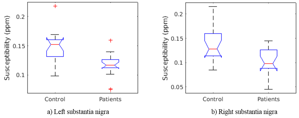
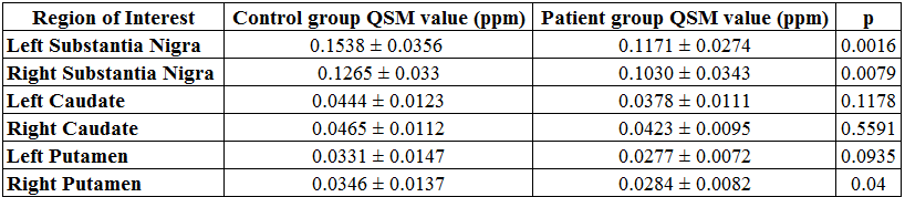
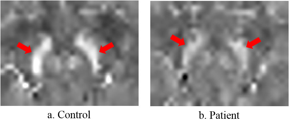
QSM reconstructions for one patient and one control are shown in figure 1. 22q11.2 DS patients had a significantly lower susceptibility than healthy controls for both, left and right substantia nigra, considering a significance value of 0.01 (figure 2). We also looked at other basal ganglia regions. Results showed no other significant differences (Table 1).
Discussion and conclusion
We here show differences in QSM measurements between subjects with the 22q11DS and healthy controls in the substantia nigra. Our result is consistent with the previous PET study showing an increased uptake of a radioligand to the dopamine transporter in patients3. While the direction of the finding might be counterintuitive, a previous study showed that QSM values and PET-DAT in the substantia nigra of healthy subjects are inversely correlated10, albeit at a trend level. Future studies will examine the relationship of this value with PET imaging in the same subjects, as well as the clinical correlation within the 22q11DS group.Acknowledgements
This publication has received funding from FONDECYT 1161448, FONDECYT 1160736, Millenium Science Initiative of the Ministry of Economy, Development and Tourism, grant Nucleus for Cardiovascular Magnetic Resonance. CONICYT Programa PIA Anillo ACT1416.References
1. Jonas, R., Montojo, C., & Bearden, C. (2014). The 22q11.2 Deletion Syndrome as a Window into Complex Neuropsychiatric Disorders Over the Lifespan. Biological Psychiatry, 75(5), 351-360. doi: 10.1016/j.biopsych.2013.07.019
2. Schneider, M., Debbané, M., Bassett, A., Chow, E., Fung, W., & van den Bree, M. et al. (2014). Psychiatric Disorders From Childhood to Adulthood in 22q11.2 Deletion Syndrome: Results From the International Consortium on Brain and Behavior in 22q11.2 Deletion Syndrome. American Journal Of Psychiatry, 171(6), 627-639. doi: 10.1176/appi.ajp.2013.13070864
3. Butcher, N., Marras, C., Pondal, M., Rusjan, P., Boot, E., & Christopher, L. et al. (2017). Neuroimaging and clinical features in adults with a 22q11.2 deletion at risk of Parkinson’s disease. Brain, 140(5), 1371-1383. doi: 10.1093/brain/awx053
4. Wang, Y., & Liu, T. (2014). Quantitative susceptibility mapping (QSM): Decoding MRI data for a tissue magnetic biomarker. Magnetic Resonance In Medicine, 73(1), 82-101. doi: 10.1002/mrm.25358
5. Pino, J., da Luz, M., Antunes, H., Giampá, S., Martins, V., & Lee, K. (2017). Iron-Restricted Diet Affects Brain Ferritin Levels, Dopamine Metabolism and Cellular Prion Protein in a Region-Specific Manner. Frontiers In Molecular Neuroscience, 10. doi: 10.3389/fnmol.2017.00145
6. Schofield, M., & Zhu, Y. (2003). Fast phase unwrapping algorithm for interferometric applications. Optics Letters, 28(14), 1194. doi: 10.1364/ol.28.001194
7. Zhou, D., Liu, T., Spincemaille, P., & Wang, Y. (2014). Background field removal by solving the Laplacian boundary value problem. NMR In Biomedicine, 27(3), 312-319. doi: 10.1002/nbm.3064
8. Milovic, C., Bilgic, B., Zhao, B., Acosta-Cabronero, J., & Tejos, C. (2018). Fast nonlinear susceptibility inversion with variational regularization. Magnetic Resonance in Medicine, 80(2), 814-821. doi: 10.1002/mrm.27073
9. Xiao, Y., Fonov, V., Chakravarty, M., Beriault, S., Al Subaie, F., & Sadikot, A. et al. (2017). A dataset of multi-contrast population-averaged brain MRI atlases of a Parkinson׳s disease cohort. Data in Brief, 12, 370-379. doi: 10.1016/j.dib.2017.04.013
10. Xiao, Y., Beriault, S., Pike, G., & Collins, D. (2012). Multicontrast multiecho FLASH MRI for targeting the subthalamic nucleus. Magnetic Resonance Imaging, 30(5), 627-640. doi: 10.1016/j.mri.2012.02.006
11. Xiao, Y., Fonov, V., Bériault, S., Subaie, F., Chakravarty, M., & Sadikot, A. et al. (2014). Multi-contrast unbiased MRI atlas of a Parkinson’s disease population. International Journal Of Computer Assisted Radiology And Surgery, 10(3), 329-341. doi: 10.1007/s11548-014-1068-y
12. Ito, H., Kawaguchi, H., Kodaka, F., Takuwa, H., Ikoma, Y., & Shimada, H. et al. (2017). Normative data of dopaminergic neurotransmission functions in substantia nigra measured with MRI and PET: Neuromelanin, dopamine synthesis, dopamine transporters, and dopamine D2 receptors. Neuroimage, 158, 12-17. doi: 10.1016/j.neuroimage.2017.06.066
Figures