2790
Microstructural changes of the cortico-striatal pathway in Fabry disease: a diffusion MRI connectometry study1Computer Science Department, University of Verona, Verona, Italy, 2Advanced Biomedical Sciences Department, University “Federico II”, Naples, Italy, 3Nephrology Unit, University “Federico II”, Naples, Italy
Synopsis
Purpose
Recent evidences suggested the presence of an alteration of the extrapyramidal system in Fabry Disease (FD)[1,2], rare X-linked lysosomal storage disorders long considered to be characterized by major cerebrovascular events only[3]. In particular, an alteration of the cortico-striatal pathway has been described in these patients, with a reduced functional connectivity between the motor cortex and the striatum, bilaterally[1]. Although a widespread alteration of the microstructural integrity of white matter (WM) has been described in FD patients[4,5,6], to date no direct investigation of the possible damage of the cortico-striatal fibers has been performed. Given this knowledge, the aim of the present work was to study the microstructural integrity of the cortico-striatal connections in FD patients, compared to a group of healthy controls (HC), to investigate the possible presence of structural connectivity changes in these connections, expanding the current knowledge about motor involvement in FD.Methods
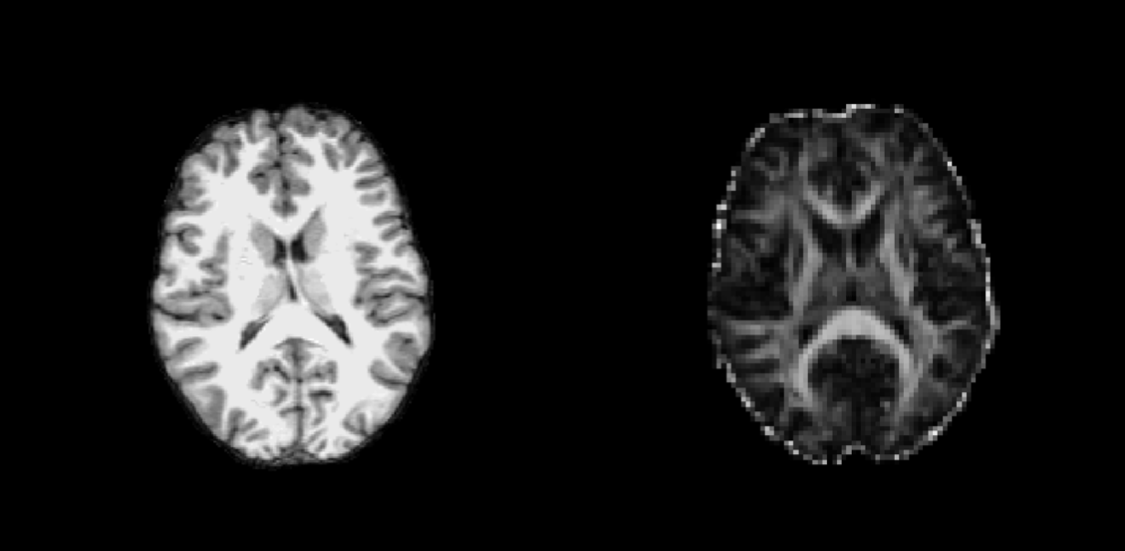
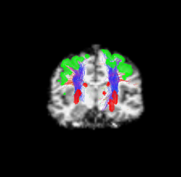
In this study, 15 FD patients (M/F=14/11, mean age $$$43.6\pm9.5$$$ years), and 14 HC (M/F=12/12, mean age $$$439\pm12.3$$$ years) were enrolled. All subjects underwent MRI scan on the same 3T scanner (Trio, Siemens Medical Systems, Erlangen, Germany). Diffusion Tensor Images (DTI) were acquired with a voxel size of $$$2.2×2.2×2.2mm^3$$$, 64 directions with $$$b-value=1000s/mm^2$$$ and nine $$$b=0s/mm^2$$$. Along with the DTI, a 3D Fluid Attenuated Inversion Recovery (FLAIR) sequence and a 3D T1-weighted volume, both with a voxel size of $$$1x1x1mm^2$$$, were also acquired. From the FLAIR images, WM lesions (when present) were segmented using a semi-automated method, and T1-weighted images were accordingly filled. DTI images were then corrected for motion and eddy currents and maps of Fractional Anisotropy (FA, reflecting fibers integrity) were computed for each subject using MRtrix3 (Figure 1). Anatomically-Constrained Tractography (ACT)[7] with iFOD2 algorithm[8] was performed to obtain 1million streamlines (Figure 2). The connectomes were built using the standard Desikan-Killiany atlas[9], implemented in FreeSurfer. For each subject, FA maps and connectomes were combined to carry on diffusion MRI connectometry[10]. Values corresponding to bundles connecting the precentral gyrus (PreCG) with the striatum (namely, caudate nucleus (CN) and putamen (PUT) were finally extracted, and a Generalized Linear Model was employed to compare the two groups, with age and gender added as covariates.Results
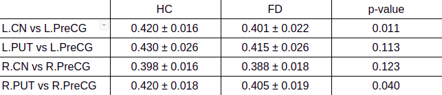
Results of the analysis are summarized in Table 1, where mean values and standard deviations for each group are reported, along with the corresponding p-values. Briefly, we proved a global reduction of FA mean values, bilaterally, along the bundles of the cortico-striatal pathway in FD patients compared to HC. In particular, we found a significant reduction mean FA values between the left PreCG and the ipsilateral CN ($$$0.420\pm0.016$$$ vs $$$0.401\pm0.022$$$ for HD and FD, respectively, $$$p=0.011$$$), as well as between tight PreCG and the ipsilateral PUT ($$$0.420\pm0.018$$$ vs $$$0.405\pm0.019$$$ for HD and FD, respectively, $$$p=0.040$$$), while the remaining connections showed similar higher mean FA values in HC compared to FD, although not reaching a statistical significance.Discussion
In this preliminary study we confirmed the presence of an alteration of the extrapyramidal system in FD patients, in line with the recent evidences suggesting the presence of widespread brain changes as a possible reflection of the subtle motor symptoms present in this condition. In particular, our result confirms the hypothesis, and expand the knowledge, about the involvement of the cortico-striatal system in this condition, showing that along with functional changes[1], microstructural damage of this pathway is present in FD patients. Previous neuroimaging studies have shown how the presence of these brain changes in FD could reflect data reported in early Parkinson's Disease (PD) patients[1,2]. Interestingly, some Authors showed the presence of an increased mean diffusivity, but no FA changes, in the cortico-striatal connections of PD patients with freezing of gait (FOG) compared with those without FOG[12]. In this light, it is noteworthy to mention that FOG, although classically considered as common in advanced subjects[11], has been also reported in a small percentage of early PD patients[13]. It is possible therefore to speculate that these microstructural changes affecting the cortico-striatal pathway in FD could somehow mirror a phenomenon also present in early PD patients, although this speculation needs to be tested and confirmed in a larger and representative group of FD patients.Conclusions
In conclusion, our preliminary results confirm the presence of an extrapyramidal involvement in FD patients, showing the presence of microstructural changes affecting the cortico-striatal pathway in this condition. These results need however to be confirmed in a larger and more representative group of patients, in order to further confirm the presence of a deep and complex involvement of motor circuits in FD.Acknowledgements
No acknowledgement found.References
1 - Cocozza S, Pisani A, Olivo G et al (2017) Alterations of functional connectivity of the motor cortex in Fabry disease: an RS-fMRI study. Neurology 88:1822–1829
2 - Russo C, Pontillo G, Pisani A et al (2018) Striatonigral involvement in Fabry disease: a quantitative and volumetric magnetic resonance imaging study. Parkinsonism Relat Disord S1353–8020:30314–30316
3 - Germain DP (2010) Fabry disease. Orphanet J Rare Dis 5:30
4 - Albrecht J, Dellani PR,Müller MJ et al (2007) Voxel based analyses of diffusion tensor imaging in Fabry disease. J Neurol Neurosurg Psychiatry 78:964–969
5 - Paavilainen T, Lepomäki V, Saunavaara J et al (2013) Diffusion tensor imaging and brain volumetry in Fabry disease patients. Neuroradiology 55:551–558
6 - Cocozza S, Pontillo G, Quarantelli M et al (2018) Default mode network modifications in Fabry disease: a resting-state fMRI study with structural correlations. Hum Brain Mapp 39:1755–1764
7 - Smith, R. E.; Tournier, J.-D.; Calamante, F. & Connelly, A. Anatomically-constrained tractography: Improved diffusion MRI streamlines tractography through effective use of anatomical information. NeuroImage, 2012, 62, 1924-1938
8 - Tournier, J.-D.; Calamante, F. & Connelly, A. Improved probabilistic streamlines tractography by 2nd order integration over fibre orientation distributions. Proceedings of the International Society for Magnetic Resonance in Medicine, 2010, 1670
9 - Desikan, R.S., Segonne, F., Fischl, B., Quinn, B.T., Dickerson, B.C., Blacker, D., Buckner, R.L., Dale, A.M., Maguire, R.P., Hyman, B.T., Albert, M.S., Killiany, R.J., 2006. An automated labeling system for subdividing the human cerebralcortex on MRI scansintogyralbasedregions of interest. Neuroimage 31, 968-980
10 - F.C. Yeh, P.-F. Tang, W.-Y. Isaac Tseng. Diffusion MRI connectometry automatically reveals affected fiber pathways in individuals with chronic stroke. NeuroImage Clin. 2013, 2:912-921
11 - Nutt JC et al, Freezing of gait: moving forward on a mysterious clinical phenomenon. Volume 10, ISSUE 8, 734-744, August 01, 2011;
12 - Vercruysse S et al. Microstructural Changes in White Matter Associated With Freezing of Gait in Parkinson’s Disease. Mov Disord. 2015 Apr;30(4):567-76. doi: 10.1002/mds.26130. Epub 2015 Jan 16
13 - Lieberman A et al. Early Freezing of Gait: Atypical versus Typical Parkinson Disorders. Parkinson’s Disease. 2015, Article ID 951645, 5 pages
Figures