2779
Comparative Corticospinal Tract Relaxation and Diffusion MRI Measures in Amyotrophic Lateral Sclerosis1Neuroradiology, Technische Universität Dresden, Dresden, Germany, 2Neurology, Technische Universität Dresden, Dresden, Germany, 3Warren Alpert Medical School, Brown University, Providence, RI, United States
Synopsis
Amyotrophic Lateral Sclerosis (ALS) is a fatal disease that primarily affects the human primary motor system. Selective neurodegeneration leads to systemic functional motor decay. We aimed to understand the relationship between cortical degeneration and the desintegration of the related motor corticospinal tract (CST) by applying both Diffusion Tensor Imaging (DTI) and the multi-component driven equilibrium single pulse observation of T1 and T2 (mcDESPOT). We found early changes in diffusion and relaxation measures indicating WM tract degeneration secondary to cortical neurodegeneration. Besides the loss of structural integrity early alterations of the myelin characteristics indicate toward changes of its compositional condition instead of early myelin loss.
INTRODUCTION
Amyotrophic Lateral Sclerosis (ALS) is a fatal disease that primarily affects the human primary motor system. Selective neurodegeneration leads to systemic functional motor decay1. We aimed to understand the relationship between cortical degeneration and the desintegration of the related motor corticospinal tract (CST) by applying both Diffusion Tensor Imaging (DTI) and the multi-component driven equilibrium single pulse observation of T1 and T2 (mcDESPOT).MATERIAL & METHODS
Twenty-seven (27) ALS patients were recruited for the NiSALS (Neuroimaging Society in Amyotrophic Lateral Sclerosis) multi-site registry that underwent cerebral neuroimaging and quantitative MRI. Age matched control subjects (n=21) free of neurological diseases were scanned to provide a comparative dataset for quantitative MRI measures. Ages and age ranges were tested for non-significant differences.
Imaging data was acquired on a SIEMENS Verio 3T scanner. DTI was acquired using an EPI sequence and a diffusion scheme with two b/values (0/100), 64 diffusion directions and a resolution of 2 mm^3. Mean diffusivity (MD), axial (AD) and radial diffusivity (RD) and fractional anisotropy (FA) measures were calculated. We also acquired isotropic 1.7 mm^3 whole-brain mcDESPOT data using the following parameters: FLASH (TR: 5.9ms, FA = [4, 5.3, 6.6, 8, 9.3, 12, 17.3, 24]°), trueFISP (TR 5.6ms; FA = [9.8, 13.1, 16.4, 19.7, 22.9, 29.5, 42.6, 59]°) and inversion prepared FLASH (TR 5.7ms, FA = 5°, TI [450, 700] ms, PE 72). The relaxation measures T1 time and T2 time, myelin water fraction (MWF) and residence time (MRT) were retrieved.
The bilateral corticospinal tract was generated as the region-of-interest (ROI) using tractographic reconstruction by means of DSIstudio. The CST diffusion measures were acquired as results of tractographic reconstruction within the edited CST ROI. The whole brain multicomponent relaxation characteristics and myelin water measures were retrieved from an in-house analysis using the approach proposed by Deoni et al2. The selective measures (T1 and T2 time, MWF and MRT) of the individual CST were retrieved masking the resulting quantitative maps with the DSIstudio tract.
RESULTS
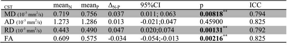
Significantly different diffusion characteristics were found in ALS patients compared to controls, in particular higher MD and lower FA indicating a general deterioration of intrinsic CST structure. Notably, RD revealed highly significant lower values in ALS CST whereas AD values were not different (see Tab. 1).
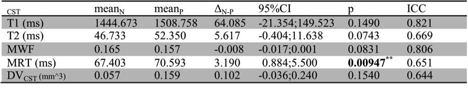
The mean CST T1 relaxation time in patients was not significantly different in ALS patients compared to controls. However, CST T2 time in patients revealed at least a trend towards significant difference with higher mean values in ALS than in controls. The mean CST MWF in ALS patients revealed lower values than in healthy controls and significantly higher values in MRT. The voxel-based approach using z-scoring3 to retrieve deficient MWF summed voxel volumes in ALS CST revealed no significant difference when compared to healthy control CST MWF (for all above see Tab.2).
DISCUSSION
Neurodegeneration of the motor system in ALS clearly involves CST integrity reflected by changes MD, RD and FA. However, the significant difference in MRT also reveals early changes in tract myelination. Whereas this might be interpreted as alteration of myelin integrity the relative myelin content measured by MWF was assumably not significantly different in ALS given the early observation at the time of disease diagnosis. Based on the results we assume that this might change in disease development.CONCLUSION
WM tract degeneration secondary to cortical neurodegeneration occurs early in disease. Besides the loss of structural integrity early alterations of the myelin characteristics indicate toward changes of its compositional condition rather than measurable early myelin loss.Acknowledgements
No acknowledgement found.References
[1] Verde et al. Arch Ital Biol. 2017 Dec 1;155(4):118-130
[2] Deoni et al., Magn Reson Med. 2008; 6:1372-87
[3] Kitzler et al. NeuroImage. 2012;59(3):2670-7
Figures