2778
Different Cortical Thinning Pattern in Primary Motor Cortex Correspond to Clinical Characteristics of Amyotrophic Lateral Sclerosis Subtypes1Department of Medical Imaging, The First Affiliated Hospital of Xi'an Jiaotong University, Xi'an, China, 2Department of Medical Imaging of the Second Hospital of Yulin, Yuling, China
Synopsis
Heterogeneity of motor phenotypes is a clinically well-recognized fundamental aspect of amyotrophic lateral sclerosis (ALS).The body region of onset is one of independent primary attributes of ALS motor phenotype heterogeneity. In order to investigate the patterns of brain atrophy between ALS patients with bulbar and limb onset and analyse its correlation with clinical characteristics , cortical thickness analyses were performed. ALS Patients with limb onset revealed the majority of significant cortical thinning in the limb segment of the motor cortex, and patients with bulbar onset, in the bulbar segments. The findings suggest that neuroimaging could be a helpful objective measure to estimate of upper motor neuron loss.
INTRODUCTION
Amyotrophic lateral sclerosis (ALS) is a rapidly progressing,phenotypically heterogeneous neurodegenerative disease characterized by deficits of the upper motor neuron (UMN) and lower motor neuron (LMN).1 Taking into account that comprehensive heterogeneity of ALS, it seems highly plausible that diverse subtypes of ALS will need diverse treatments .2 However, the mechanisms underlying neurodegeneration in ALS subtypes are not yet completely understood . A valid objective biomarker that correlated with disease pathology of ALS subtypes was likely to contribute to patients' clinical management . The body region of onset is one of independent primary attributes of ALS motor phenotype heterogeneity .3 Therefore, in order to investigate the patterns of brain atrophy between ALS patients with bulbar and limb onset and analyse its correlation with clinical characteristics , cortical thickness analyses were performed.METHODS
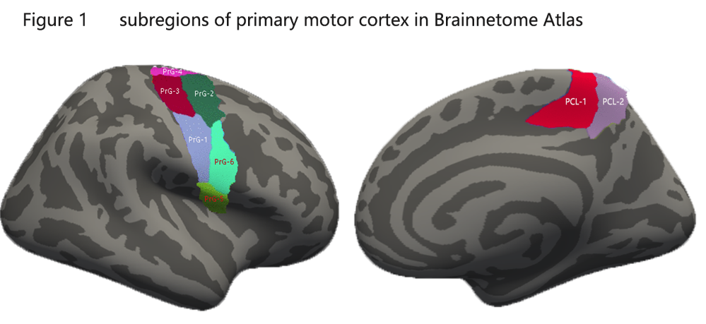
Fifty-nine ALS patients that included 39 patients with limb onset , out of which 13 with bulbar onset and 66 healthy controls were enrolled in this study. T1-weighted images of the brain were acquired on high field 3 T scanner. Surface based cortical morphology measures were performed using the FreeSurfer programme.Based on previous observations, primary motor cortex was selected as region of interest. Besides, in order to evaluate more precise brain morphometric changes, we used the Brainnetome Atlas, 4 in which primary motor cortex was segmented into 8 subregions according to different function of motor homunculus (Figure 1). Multivariates analysis of covariance (ANCOVA) with controlling age and averaged cortical thickness was conducted. Post-hoc multiple comparison with Bonferroni test was performed. The Pearson correlation analyses were used to assess relationship between cortical thickness with clinical measurements.RESULTS
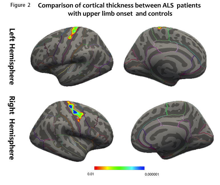
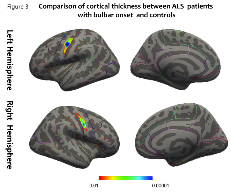
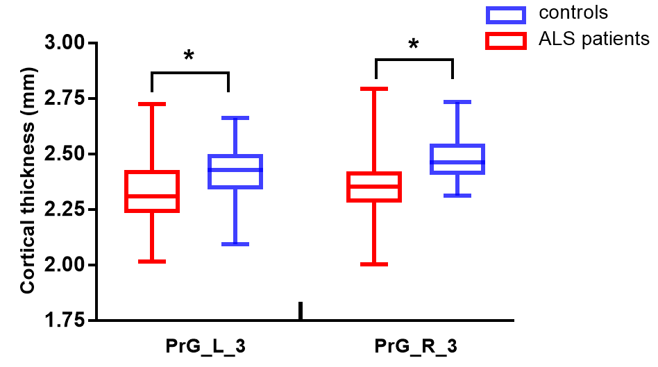
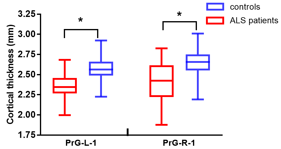
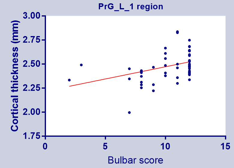
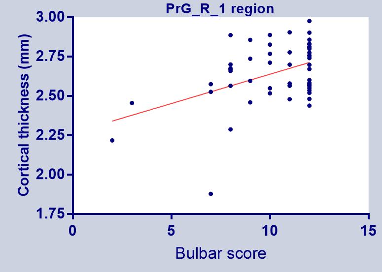
When compared to healthy controls , ALS patients confirmed significant cortical thinning ofthe bilateral PMC. ALS patients with limb onset showed the majority of significant cortical thinning in the bilateral limb segment of the motor homunculus(figure 2). On the contrary , ALS patients with bulbar onset showed the majority of significant cortical thinning in the bilateral bulbar segment of the motor homunculus(figure 3). By using the precise Brainnetome Atlas, the results were in consistent with previous vertex-wise analysis. ALS patients with limb onset showed reduced cortical thickness primarily in the bilateral PrG-3 regions, indicated function impairment in upper limb region(figure4). In contrast, ALS patients with bulbar onset showed reduced cortical thickness primarily in the bilateral PrG-1 regions, indicated function impairment in bulbar region(figure 5).The correlation analyses revealed significant positive relationship between bulbar functional subscores with bilateral PrG-1 cortical thickness of ALS patients (figure 6).
DISCUSSION
This study mainly investigated the patterns of cortical thinning between ALS patients with bulbar onset and patients with limb onset. ALS Patients with limb onset revealed the majority of significant cortical thinning in the limb segment of the motor cortex, and patients with bulbar onset, in the bulbar segments. Further more, the results of ROI-based analysis were in consistent with previous vertex-wise analysis . The research finding supports the concepts of cortical focality 3and cortical influences drive amyotrophic lateral sclerosis.5 Heterogeneity of motor phenotypes is a clinically well-recognized fundamental aspect of amyotrophic lateral sclerosis (ALS) .Different ALS-subtypes will probably respond differently to modifying therapies .2 Cortical thickness measurement appears to provide a sensitive estimate of upper motor neuron loss with the potential to be biomarkers to improve ALS precise therapy trials.CONCLUSION
Focal cortical thinning within the motor homunculus corresponds with functional disabilityin ALS, implies that neuroimaging could be a helpful objective measure to estimate of upper motor neuron loss. Cortical thinning pattern in primary motor cortex with ALS phenotype may be an useful biomarkers in clinical precise therapy trials in individual.Acknowledgements
No acknowledgement found.References
1 Brown RH, Al-Chalabi A. Amyotrophic Lateral Sclerosis. N Engl J Med 2017; 377(2): 162-172.
2 van Es MA, Hardiman O, Chio A, Al-Chalabi A, Pasterkamp RJ, Veldink JH, van den Berg LH. Amyotrophic lateral sclerosis. Lancet 2017; 390(10107): 2084-2098.
3 Ravits JM, La Spada AR. ALS motor phenotype heterogeneity, focality, and spread: deconstructing motor neuron degeneration. Neurology 2009; 73(10): 805-811.
4 Fan L, Li H, Zhuo J, Zhang Y, Wang J, Chen L, Yang Z, Chu C, Xie S, Laird AR, Fox PT, Eickhoff SB, Yu C, Jiang T. The Human Brainnetome Atlas: A New Brain Atlas Based on Connectional Architecture. Cereb Cortex 2016; 26(8): 3508-3526.
5 Eisen A, Braak H, Del TK, Lemon R, Ludolph AC, Kiernan MC. Cortical influences drive amyotrophic lateral sclerosis. J Neurol Neurosurg Psychiatry 2017; 88(11): 917-924.
Figures