2769
The diagnosis of Internet gaming disorder by MR imaging: A MRI based analysis1Institute of Science and Technology for Brain, Institute of Science and Technology for Brain-Inspired Intelligence, Fudan University, shanghai, China, 2Institute of Science and Technology for Brain-Inspired Intelligence, Fudan University, shanghai, China, 3Department of Radiology, Ren ji Hospital, School of Medicine, Shanghai Jiao Tong University, shanghai, China
Synopsis
The internet gaming disorder has become one of the most serious healthy problem among teenagers, the questionnaire and scale are widely used to IGD diagnostic. However, the underlying neural mechanism of IGD was still unclear. Current study present an evidence that cerebral morphometric alteration could be used to identified IGD from normal, and may also help for further study about IGD.
Introduction
The internet gaming disorder has become one of the most serious health problem among teenagers, the questionnaire and scale are widely used to IGD diagnostic, such as CIAS [1], YIAT [2], but the underlying neural mechanism is still unclear. To identify the cerebral radiomic features related to diagnosis of IGD, we extract the features form gray matter from T1weighted images and white matter from diffusion tensor images, considering that IGD as a psychiatric disorder, the neural imaging evidence may provide a more powerful evaluation of IGD diagnosis.Method
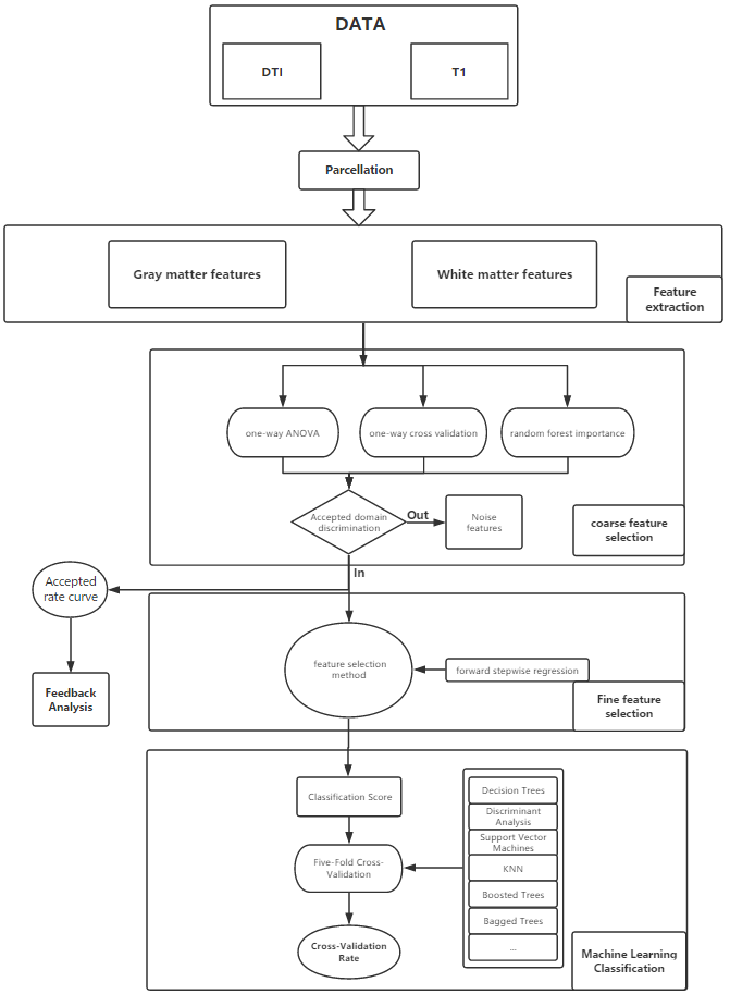
140 participants which reported playing massive multiplayer role play game (MMORPG) were recruited for current study and approved by the local ethics committee, all brain imaging data were scanned at Renji hospital in Shanghai. A modified diagnostic questionnaire of Young’s Diagnostic Questionnaire for internet addiction with 8 items and Chen Internet Addiction Scale (CIAS) with 26 items were used for the diagnosis of IGD. 59 participants were finally determined as IGD. The T1-weighted images were processed with Freesurfer (recon-all processing pipeline with the Desikan-Killiany-Tourville atlas) and Mindboggle to generate the features of each label. Totally 1333 shape-related features of gray matter were extracted from each T1-weighted images. Distribution metrics (mean, standard deviation, skew, kurtosis) were extracted from each shape properties (local thickness, mean curvature, convexity, geodesic depth, and travel depth) in labeled region. The diffusion tensor images were processed mainly by FSL, the process pipeline including eddy current correction, brain extraction and tensor model fitting. Distribution metrics (mean, standard deviation, skew and kurtosis) were extracted from each diffusion parameter maps (fractional anisotropy, mean diffusivity, axial diffusivity, and radial diffusivity) for each label with the JHU-ICBM-81 in participant individual space. totally 768 features were extracted for represent the properties of white matter. Totally 2101 features of each participant were extracted for further classification. Forward stepwise regression was firstly used to select the features to generate classifier, all of the selected features were then embedded in a repeated k-fold (k=5) cross validation framework to obtain unbiased estimates of classification error (see Fig 1).Results
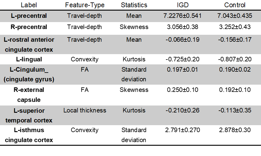
In the construction of the classifier to discriminate between IGD and health controls, the support vector machine was selected with average accuracy of 85.7%, sensitivity 86.8%, and specificity 85.1%. All of the selected features include the mean travel depth of left precentral cortex, mean travel depth of left rostral anterior cortex, travel depth skewness of right precentral, convexity standard deviation of left isthumus cingulate cortex,convexity kurtosis of left lingual,local thickness kurtosis of left superior temporal, FA standard deviation of left cingulum, and FA skewness of right external capsule. (See Fig 2).
Discussion
The mainly finding of current study was that brain imaging parameter could be used for discrimination of IGD from healthy. The alteration of cortical shape in anterior cingulate cortex was significant associate with the inhibitory control ability and perform an important role in processing addiction related stimuli [3, 4]. The precentral was involved in most of motor related functions[5], and suggest the importance of alteration in attentional bias and motor ability after long time use of internet game. Compare to the previous studies with widespread change of white matter integrity in group level comparison, our research identified local alteration in the skew of FA in cingulum which connect the medial temporal lobe and posterior cingulate regions[6] and skew of FA in external capsule which connect the anterior and posterior attentional system[7]. These finding may suggest an alteration of attentional function and default mode network function. Our finding indicates an effective classification between IGD and healthy, and an automatic pipeline could also be easily applied in clinically diagnose.Acknowledgements
No acknowledgement found.References
1. Ko, C.H., et al., Screening for Internet addiction: an empirical study on cut-off points for the Chen Internet Addiction Scale. Kaohsiung Journal of Medical Sciences, 2005. 21(12): p. 545-551.
2. Young, K.S., Internet Addiction: The Emergence of a New Clinical Disorder. Cyberpsychology & Behavior, 2009. 1(3): p. 237-244.
3. Goldstein, R.Z., et al., Role of the anterior cingulate and medial orbitofrontal cortex in processing drug cues in cocaine addiction. Neuroscience, 2007. 144(4): p. 1153-1159.
4. Lin, F., et al., Abnormal White Matter Integrity in Adolescents with Internet Addiction Disorder: A Tract-Based Spatial Statistics Study. Plos One, 2012. 7(1): p. e30253.
5. Desmurget, M., et al., Neural representations of ethologically relevant hand/mouth synergies in the human precentral gyrus. Proc Natl Acad Sci U S A, 2014. 111(15): p. 5718-5722.
6. Zhang, Y., et al., Diffusion tensor imaging of cingulum fibers in mild cognitive impairment and Alzheimer disease. Neurology, 2007. 68(1): p. 13.
7. Pastura, G., et al., Exploratory analysis of diffusion tensor imaging in children with attention deficit hyperactivity disorder: evidence of abnormal white matter structure. Attention Deficit & Hyperactivity Disorders, 2015. 8(2): p. 65-71.
Figures