2766
Sexual dimorphism in the young adult brain using magnetic resonance imaging: The effect of the field strengths1Department of Biomedical Imaging and Radiological Science, China Medical University, Taichung, Taiwan, 2Department of Radiology, China Medical University Hospital, Taichung, Taiwan, 3Department of Radiology, Taichung Tzu Chi Hospital, Taichung, Taiwan
Synopsis
The existing reports regarding sexual dimorphism in brain structures are not confluent and generally heterogeneous. MRI instrument-related factor such as field strength is one of the greatest contributors to the brain quantification variability, but its effect on sexual dimorphism in brain structures remains unclear. In this study, we found that due to the image contrast differences arising from differences in field strengths, the sex dimorphism in brain morphology appears to exist dependent of field strengths. It suggests field strength should be considered as one important factor that contributes to the inconsistency in the sex dimorphism in brain across literatures.
Introduction:
Sex-related differences in human brain structures have now gained increasing attention in a number of investigations.1, 2 The existing reports in literature are not confluent and generally heterogeneous. Possible reasons for the discrepancy can be explained by the varying sample sizes, age range, and the processing techniques.1 For the MRI-based quantitative characterization of the human brain, MRI instrument-related factor such as field strength is one of the greatest contributors to the brain quantification variability.3 However, whether the discrepancy of sexual dimorphism in brain structures varies as a function of field strengths remains unclear. Therefore, the central goal of this study was to investigate the potential effect of field strengths on sex-related differences in the brains of a young cohort aged 20-30 years in terms of both global and regional analysis. The narrow age range has a favorable effect on removing the age-related pathology and therefore truly reflects the sex differences in mature brains.Methods:
Study design: A total of 60 healthy subjects (27 females, 23.5 ± 2.5 years old) were recruited in this study. Informed consent was obtained using IRB-approved protocol. All volunteers were scanned at both 1.5T (GE, Signa, Excite HDx, Wisconsin, USA) and 3T (GE, Signa, Excite HDxt, Wisconsin, USA) scanners on the same day. The order of 1.5T or 3T scanning was randomized and separated by at least 30 min. MRI measurement: For the 1.5T scanner, the axial T1W image was obtained with fast spoiled gradient echo (FSPGR). The scanning parameters are TR/TE/FA=6.22 msec/1.99 msec/12°, and time of inversion (TI)=450 msec. For the 3T scanner, T1W image was acquired axially using FSPGR as well. The scanning parameters are TR/TE/FA=8.02 msec/2.99 msec/12°, and TI=450 msec. Both sequences had a spatial resolution=1x1x1mm3 and number of slices=170. Data analysis: The region of interest of whole brain gray matter (GM) volume from the T1W image was delineated by FSL software for each subject. Segmentation of regional brain structures from T1W image and estimation of regional volumes were performed using the MRIcloud.4 For the voxel-based morphometry (VBM) analysis, DARTEL toolbox was performed with SPM8 and Matlab by the default settings. Within the framework of the general linear model (GLM), the sexual dimorphism in the brain was analyzed by SPM8 after controlling for age, GM volume, handedness, and body mass index (BMI) effects.Results:
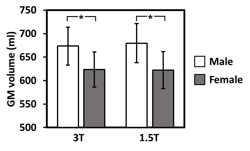
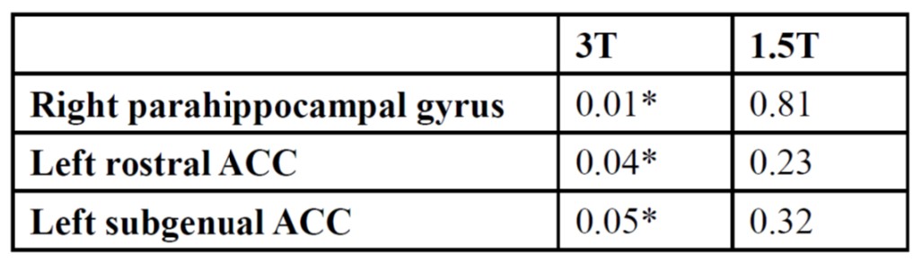
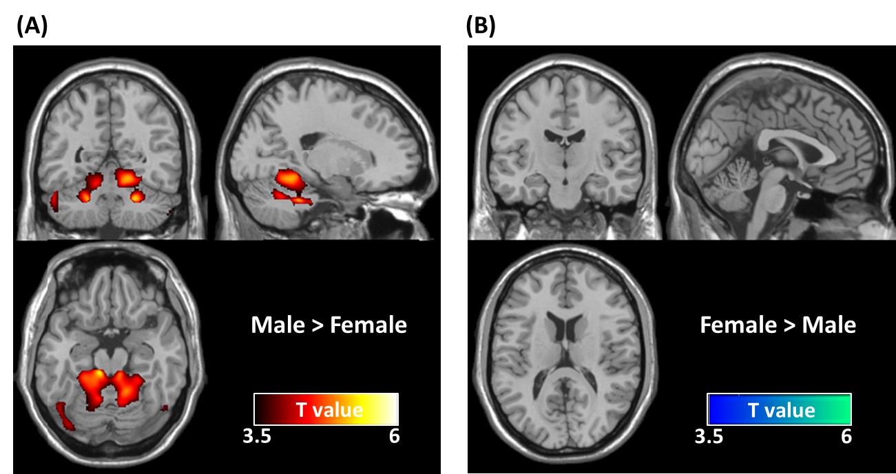
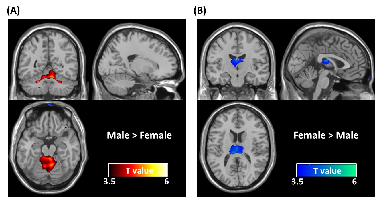
Global GM volumetric measures: Comparisons of global GM volumes between male and female subjects are shown in Fig. 1. Males had larger raw volumes of GM than their female counterparts at both field strengths, and the field strength had an insignificant effect on GM volume measurements (P>0.05 for both sexes). Comparison of regional brain volumes: Using the MRIcloud4 atlas-based parcellation approach, males had significantly larger regional brain volumes in right parahippocampal gyrus, left rostral anterior cingulate cortex (ACC), and left subgenual ACC than females (all P <0.05) at 3T. However, no sex-related differences in regional brain volumes were detected at 1.5T (Fig. 2). The opposite effect of females with larger regional brain volumes than males was not observed at both field strength. Voxel-wise differences in GM: VBM results of voxel-wise differences in brain volume are presented in Fig. 3 and Fig. 4 for 3T and 1.5T, respectively. Sexual dimorphism in cerebellum was detected at both field strengths, with cerebellar volume larger in males when compared to that in females. Females showed more GM in thalamus, but this effect was only significant at 1.5T.Discussion and Conclusion:
In this study, we explored the sex-related differences in human brain morphology in global as well as regional GM in a representative young-aged population within a limited range of age. Sex dimorphism in GM was observed not only globally but also regionally. However, the sex dimorphism in brain morphology appears to exist dependent of field strengths. It has been recognized that the image contrast differences arising from differences in field strengths, and the brain volume estimations could also be field-strength dependent.5 Therefore, MRI-instrument specific factor such as field strength should be considered as one of the important factors that contributes to the inconsistency in the sex dimorphism in human brain across literatures.Acknowledgements
No acknowledgement found.References
1. Chen X, Sachdev PS, Wen W, Anstey KJ. Sex differences in regional gray matter in healthy individuals aged 44-48 years: a voxel-based morphometric study. NeuroImage 2007; 36(3): 691-9.
2. Kruggel F. MRI-based volumetry of head compartments: normative values of healthy adults. NeuroImage 2006; 30(1): 1-11.
3. Han X, Jovicich J, Salat D, van der Kouwe A, Quinn B, Czanner S et al. Reliability of MRI-derived measurements of human cerebral cortical thickness: the effects of field strength, scanner upgrade and manufacturer. NeuroImage 2006; 32(1): 180-94.
4. Tang X, Oishi K, Faria AV, Hillis AE, Albert MS, Mori S et al. Bayesian Parameter Estimation and Segmentation in the Multi-Atlas Random Orbit Model. PloS one 2013; 8(6): e65591.
5. Jovicich J, Czanner S, Han X, Salat D, van der Kouwe A, Quinn B et al. MRI-derived measurements of human subcortical, ventricular and intracranial brain volumes: Reliability effects of scan sessions, acquisition sequences, data analyses, scanner upgrade, scanner vendors and field strengths. NeuroImage 2009; 46(1): 177-92.
Figures