2763
Brain gray matter correlates of extraversion: A systematic review and meta-analysis of voxel-based morphometry studies1Huaxi MR Research Center (HMRRC), Chengdu, China
Synopsis
Extraversion is a fundamental personality dimension closely related to individuals’ physical and mental health. Although increasing studies have attempted to identify the neurostructural markers of extraversion but have yielded inconsistent and heterogeneous results. The current study aims to reach a comprehensive understanding of brain gray matter (GM) correlates of extraversion by using a systematic review and meta-analysis approach. Our review revealed a preliminary outline of the brain GM differences related to extraversion in distributed brain regions. Our meta-analysis of voxel-based morphometry studies identified six core brain regions correlated with extraversion and revealed the potential effect of gender and age.
Introduction
Extraversion is one of the core dimensions of personality in virtually all personality theories 1-7, which is closely related to individuals’ physiological and psychological health. With the rapid development in personality neuroscience in the past decade, an increasing number of neuroimaging studies have been conducted to uncover how extraversion may be related to individual differences in brain gray matter (GM) structure. However, the findings of these studies are inconsistent and heterogeneous. In this research, we performed a systematic review and meta-analysis to achieve a comprehensive understanding of brain GM correlates of extraversion.Methods
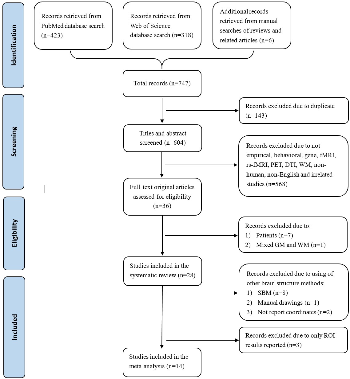
A systematic literature search and data selection were performed according to the Preferred Reporting Items For Systematic Reviews and Meta-Analyses (PRISMA) statement 8 (see Fig. 1). As a consequence, a total of 28 studies were included in the systematic review, and a total of 14 studies were entered into the meta-analysis that enrolled 1,547 healthy subjects (857 females, 33.8 ± 7.3 years old), and covered 100 peaks stereotaxic coordinates. Basical information of each identified study was described and categorized in the review. To detect brain regions whose GMV is stablely correlated with extraversion, a meta-analysis was conducted using anisotropic effect-size seed-based d mapping (AES-SDM) software. To obtain more stable and reliable results of meta-analysis, we set permutation at 20 9 and used stantard SDM thresholds (voxel-wise p < 0.005, SDM-Z > 1 and cluster size > 10 voxels), which have been verified to effectively balance false positive and negative 10-13. Moreover, heterogeneity analyses with Q statistics and jackknife sensitivity analyses were performed to examine inter-study heterogeneity and reliability of the findings respectively. Meta-regression analyses were carried out to investigate the potential effect of gender and age with more stringent thresholds (voxel-wise p < 0.0005, z > 1 and cluster size > 10 voxels) to decrease probability of false findings 11.Results
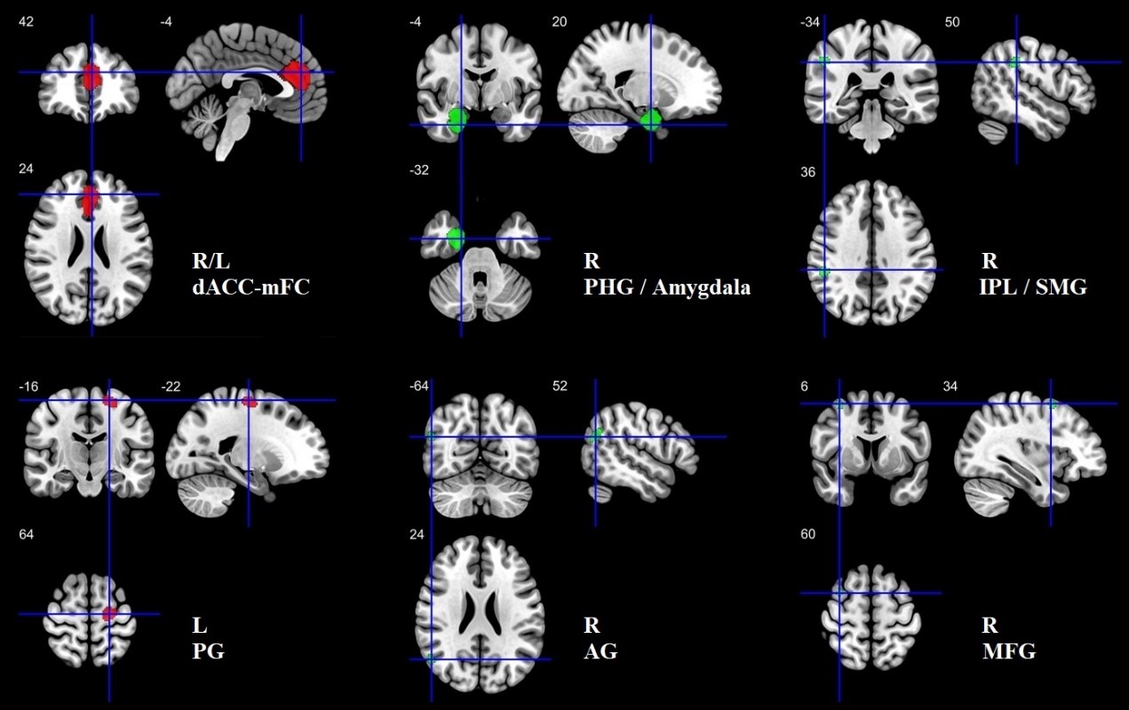
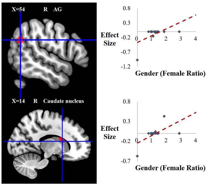
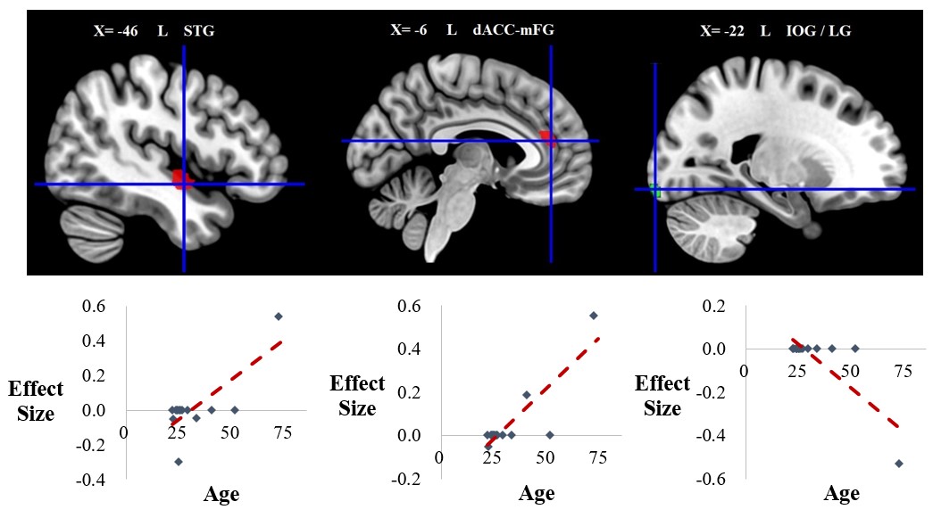
The systematic review showed the heterogeneity of the previous findings regarding the GM correlates underlying extraversion. Moreover, the inconclusive neuroanatomical correlates of extraversion might be largely due to the differences in sample size, personality measurements, morphometrical techniques, nuisance covariates, and etc. The meta-analysis revealed six core brain clusters significantly associated with extraversion including bilateral dorsal anterior cingulate cortex-medial frontal gurys (dACC-mFG), left precentral gyrus (PG), right parahippocampal gyrus/Amygdala (PHG/Amygdala), right inferior parietal lobule/supramarginal gyrus (IPL/SMG), right angular gyrus (AG) and right middle frontal gyrus (MFG), the first two of which were identified to have positive correlations, and the last four of which were identified to have negative correlations (see Fig. 2). Furthermore, the current results showed obvious hemispheric asymmetry in the GM correlates of extraversion. Specifically, the positive correlations were mostly found in the left hemisphere whereas the negative correlations were largely found in the right hemisphere. Jacknife sensitive analyses revealed that all six brain clusters were repeatedly identified in no less than twelve combinations, suggesting that the results observed in main meta-analysis were highly consistent. Heterogeneity analyses exhibited a mount of brain regions which had significant inter-study variabilities, consistent with the findings in systematic review. To account for the heterogeneity between studies, we performed meta-regression analyses to explore potential effect of gender and age. The meta-regression analysis of gender showed a gender difference in the right AG and the right caudate nucleus (see Fig. 3). The meta-regression analysis of age indicated a age effect in the left superior temporal gyrus (STG), the left dACC-mFG and the left inferior occipital gyrus/ lingual gyrus (IOG/LG) (see Fig. 4).Discussion
Despite the findings of the previous studies included in our systematic review were relatively heterogeneous, a preliminary outline of the brain GM differences linked to extraversion was delineate in distributed brain regions widespread in frontal, temporal, parietal, occipital and subcortical structures. Moreover, our meta-analysis revealed inter-study convergence in dACC-mFG, PG, PHG/Amygdala, IPL/SMG, AG and MFG, consistent with the findings of fMRI studies during emotional process, reward learning and social cognition14-17. Our findings are also in line with studies in patients reporting that brain GM loss in these regions are correlated with reduced extraversion 18. Additionally, the gender and age effects observed in current study may explain the heterogeneity across studies and hint the importance of covariates selection for model.Conclusion
In brief, based on a systematic review and meta-analysis, the current study provided a preliminary brain GM map of extraversion, identified six core regions linked to extraversion and further showed gender and age effect on extraversion-GM associations. To our knowledge, this is the first study to reveal a comprehensive picture of brain GM correlates of extraversion.Acknowledgements
No acknowledgement found.References
1. Depue RA, Collins PF. Neurobiology of the structure of personality: dopamine, facilitation of incentive motivation, and extraversion. The Behavioral and brain sciences. 1999;22:491-517; discussion 518-469.
2. Goldberg LR. An alternative "description of personality": the big-five factor structure. J Pers Soc Psychol. 1990;59:1216-1229.
3. Gray JA. The psychophysiological basis of introversion-extraversion. Behaviour research and therapy. 1970;8:249-266.
4. Eysenck HJ: The biological basis of personality, Charles C. Thomas, Spring-field, IL; 1967.
5. Tellegen A: Multidimensional personality questionnaire manual. University of Minnesota Press; 1982.
6. Cattell RB, Eber HW, Tatsuoka MM. Handbook for the sixteen personality questionnaire (16PF). Institute for Personality and Ability Testing. 1980.
7. Cloninger CR, Przybeck TR, Svrakic DM. The tridimensional personality questionnaire: US normative data. Psychological reports. 1991;69:1047-1057.
8. Liberati A, Altman DG, Tetzlaff J, Mulrow C, Gotzsche PC, Ioannidis JP, Clarke M, Devereaux PJ, Kleijnen J, Moher D. The PRISMA statement for reporting systematic reviews and meta-analyses of studies that evaluate healthcare interventions: explanation and elaboration. BMJ (Clinical research ed). 2009;339:b2700.
9. Yao YW, Liu L, Ma SS, Shi XH, Zhou N, Zhang JT, Potenza MN. Functional and structural neural alterations in Internet gaming disorder: A systematic review and meta-analysis. Neurosci Biobehav Rev. 2017;83:313-324.
10. Han JE, Boachie N, Garcia-Garcia I, Michaud A, Dagher A. Neural correlates of dietary self-control in healthy adults: A meta-analysis of functional brain imaging studies. Physiology & behavior. 2018;192:98-108.
11. Pan P, Zhan H, Xia M, Zhang Y, Guan D, Xu Y. Aberrant regional homogeneity in Parkinson's disease: A voxel-wise meta-analysis of resting-state functional magnetic resonance imaging studies. Neurosci Biobehav Rev. 2017;72:223-231.
12. Radua J, Mataix-Cols D, Phillips ML, El-Hage W, Kronhaus DM, Cardoner N, Surguladze S. A new meta-analytic method for neuroimaging studies that combines reported peak coordinates and statistical parametric maps. European psychiatry : the journal of the Association of European Psychiatrists. 2012;27:605-611.
13. Yang X, Tian F, Zhang H, Zeng J, Chen T, Wang S, Jia Z, Gong Q. Cortical and subcortical gray matter shrinkage in alcohol-use disorders: a voxel-based meta-analysis. Neurosci Biobehav Rev. 2016;66:92-103.
14. Canli T, Amin Z, Haas B, Omura K, Constable RT. A double dissociation between mood states and personality traits in the anterior cingulate. Behav Neurosci. 2004;118:897-904.
15. Canli T, Zhao Z, Desmond JE, Kang E, Gross J, Gabrieli JD. An fMRI study of personality influences on brain reactivity to emotional stimuli. Behav Neurosci. 2001;115:33-42.
16. Hooker CI, Verosky SC, Miyakawa A, Knight RT, D'Esposito M. The influence of personality on neural mechanisms of observational fear and reward learning. Neuropsychologia. 2008;46:2709-2724.
17. Hassabis D, Spreng RN, Rusu AA, Robbins CA, Mar RA, Schacter DL. Imagine all the people: how the brain creates and uses personality models to predict behavior. Cereb Cortex. 2014;24:1979-1987.
18. Mahoney CJ, Rohrer JD, Omar R, Rossor MN, Warren JD. Neuroanatomical profiles of personality change in frontotemporal lobar degeneration. Br J Psychiatry. 2011;198:365-372.
Figures