2762
Acute and Chronic intranasal oxytocin differentially affect brain functional connectivity1Center for Neuroscience and Cognitive System, Istituto Italiano di tecnologia, Rovereto, Italy, 2Centro Interdipartimentale Mente/Cervello (CIMeC), University of Trento, Rovereto, Italy
Synopsis
Intranasal oxytocin (OXT) administration has shown promise as a putative treatment for disorders characterized by social impairments. However, the brain-wide substrates engaged by this neuropeptide remain elusive. By using mouse fMRI, we show that the circuits engaged by intranasal OXT are differentially affected by the duration of OXT dosing. Specifically, acute OXT administration increases brain connectivity in key nodes of the social brain. By contrast, repeated dosing exacerbates inter-regional coupling and results in paradoxical social impairments in control “wild type” mice. These result have implications for clinical testing of OXT in control and pathological conditions.
Introduction
Oxytocin (OXT), a neuropeptide implicated in the modulation of pro-social and cognitive functions1,2 has been proposed as a treatment for disorders characterized by social impairments, such as autism and schizophrenia3,4.We recently showed that acute intranasal dosing of OXT robustly activates several functional networks involved in social and affective behavior in rodents6. However, the therapeutic use of this drug requires repeated dosing5 and previous animal studies have revealed diverging behavioral effects of OXT when administered acutely or chronically in rodents7. Here we test the hypothesis that OXT dosing regimen result in differential neuro-functional and behavioral effects via recruitment of different functional connectivity networks. To test this hypothesis, we mapped the regional fMRI response and functional connectivity produced by acute and chronic intranasal administration of OXT in the mouse.Methods
Intranasal OXT (0.3 IU) or saline were intranasal administered to adult male C57Bl6/J mice twice a day, for 7 consecutive days7. On day 7, 1 hour later than the last administration, mice were tested in a male-female social interaction test, in order to evaluate the effect of chronic OXT dosing on social and communicative responses. On the following day (day 8), mice underwent CBV-weighted fMRI mapping after an intranasal OXT challenge (0.8 IU)6. We mapped connectivity using between-subject seed-based correlation maps obtained via group-level mapping of functional covariance8, using FSL randomize non parametric permutation testing9, and employing a cluster correction for family-wise error rate (FWER).Results
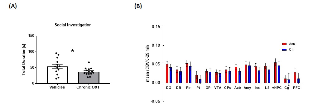
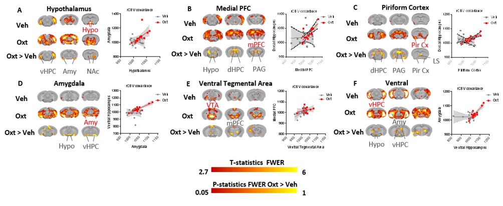
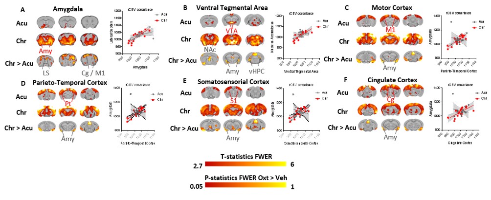
Recapitulating previous reports, chronic administration of OXT resulted in reduced social communication, as assessed with a male-female social interaction (Fig.1A). Interestingly, this effect was not associated with a differential response in the regional magnitude of the CBV-weighted fMRI response elicited by intranasal OXT dosing (Fig.1B). We therefore tested whether inter-regional connectivity (as opposed to the magnitude of BOLD response) could explain the observed behavioural differences. Acute OXT administration in OXT-naïve animals promoted inter-regional connectivity between cortico-limbic, olfactory and hippocampal areas (Fig.2) that are part of rodent social brain. Chronic OXT administration resulted in a stronger functional coupling between these regions, plus a non-canonical coupling between forebrain social areas, and large neo-cortical regions (Fig.3).Discussion
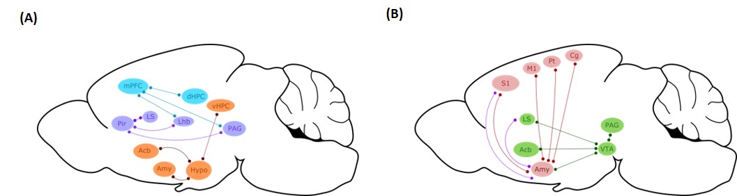
Our results show that exogenous OXT administration increases brain connectivity within the rodent social brain, and that extended dosing leads to a non-canonical inter-regional coupling between the amygdala and large neo-cortical areas that are typically not involved in socially-relevant behaviours (Fig.4). Importantly, we found this latter connectivity fingerprint to be associated with impaired social recognition in wild type mice. We propose that the atypical recruitment of large neocortical motor-sensory areas may underlie an abnormal gating of different sensorial inputs by the amygdala, biasing social responses in rodents10. From a mechanistic standpoint, it is possible that overstimulation of endogenous OXT synthesis mediated by the exogenous OXT administration could result in the functional recruitment of long-range substrates enriched with oxytocin receptors11, leading to a hyper-synchronized coupling between these brain regions. While socially detrimental in the healthy brain, further investigations needed to address if the resulting hyper-connected state could be therapeutically effective in restoring reduced functional connectivity associated with developmental connectopathies such as autism spectrum disorders and schizophrenia.Conclusions
We provide neuroimaging evidence that acute and chronic OXT differentially affect interregional connectivity. The observed functional divergences should be taken into consideration in the design and interpretation of human and animal studies of the substrates and mechanism engaged by exogenously administered OXT.Acknowledgements
No acknowledgement found.References
References
1. Insel TR. The challenge of translation in social neuroscience: a review of oxytocin, vasopressin, and affiliative behavior. Neuron 2010; 65: 768-779.
2. Young LJ. When too much of a good thing is bad: chronic oxytocin, development, and social impairments. Biol Psychiatry 2013; 74(3):160-1.
3. Meyer-Lindenberg A, Domes G, Kirsch P, Heinrichs M. Oxytocin and vasopressin in the human brain: social neuropeptides for translational medicine. Nat Rev Neurosci 2011;12: 524-538.
4. Young LJ and Barrett CE. Neuroscience. Can oxytocin treat autism? Science 2015;347 (6224):825-6.
5. Macdonald K and Feifel D. Helping oxytocin deliver: considerations in the development of oxytocin-based therapeutics for brain disorders. Front Neurosci. 2013;7:35.
6. Galbusera A, De Felice A, Girardi S et al. Intranasal Oxytocin and Vasopressin Modulate Divergent Brainwide Functional Substrates. Neuropsychopharmacology. 2017;42(7):1420-1434.
7. Huang H, Michetti C, Busnelli M et al. Chronic and acute intranasal oxytocin produce divergent social effects in mice. Neuropsychopharmacology. 2014;39(5):1102-14.
8. Pagani M, Bifone A and Gozzi A. Structural covariance networks in the mouse brain. Neuroimage. 2016 Apr 1;129:55-63.
9. Mechelli, A., K. J. Friston, R. S. Frackowiak and C. J. Price. Structural covariance in the human cortex. J Neurosci 2005; 25(36):8303-10.
10. Chen P and Hong W. Neural Circuit Mechanisms of Social Behavior. Neuron. 2018;98(1):16-30.
11. Ludwig M and Leng G. Dendritic peptide release and peptide-dependent behaviours. Nat Rev Neurosci. 2006;7(2):126-36.
Figures