2761
Medial temporal cortical changes in response to yoga and aerobic exercise interventions in early psychosis patientsMelissa L Woodward1, Jingxia Lin2, Wayne Weizhong Song3, William G Honer3, Eric YH Chen2, and Donna J Lang1
1Radiology, University of British Columbia, Vancouver, BC, Canada, 2Psychiatry, University of Hong Kong, Hong Kong, China, 3Psychiatry, University of British Columbia, Vancouver, BC, Canada
Synopsis
Early psychosis patients exhibit cortical reductions and poor cardiovascular health, which may be worsened by antipsychotic medication. Aerobic exercise and yoga may be able to remediate cortical loss and improve symptom severity. First-episode psychosis patients who completed a twelve-week exercise program showed increased cortical volume and thickness compared to waitlist controls with differential effects of aerobic exercise and yoga. Exercise-mediated changes in brain measures were associated with greater improvement in symptom severity scores. Both aerobic exercise and yoga may have neuroanatomical and clinical benefits for early psychosis patients and may be a safe, cost-effective adjunct treatment.
Introduction
Psychosis patients exhibit neuroanatomic deficits even at early stages of illness.1 Decreased hippocampal volume and reduced cortical thickness is common, and antipsychotic medications may further reduce cortical gray matter.2 Concomitant cardiovascular disease is common and serves as the primary contributor to premature mortality for patients with schizophrenia.3 Antipsychotic medications may further reduce cortical gray matter and worsen cardiovascular and metabolic symptoms.4,5 Physical activity may be a key adjunct treatment for patients following their first psychotic episode to counteract cardiovascular concerns. Exercise may remediate neuroanatomic deficits by triggering the release of neuronal growth factors in the hippocampus and other regions.6,7 The potential for hippocampal changes to extend to adjacent cortical areas in schizophrenia is unclear. Medial temporal cortical regions (entorhinal, fusiform, and parahippocampal cortex) serve as the intermediary between the hippocampus and the frontal cortex (see Figure 1). Assessing the impact of exercise of these regions, and the orbitofrontal cortex will provide insight into the potential for exercise-induced remediation beyond the hippocampus. Different types of exercise may confer varying benefits as the majority of research showing positive brain changes has involved aerobic exercise. Yoga has previously been shown to provide neurocognitive benefits to first-episode psychosis patients but its ability to contribute to structural brain changes requires further investigation.8Methods
74 female first-episode psychosis patients were recruited from three hospital sites in Hong Kong. Participants were randomized into yoga, aerobic exercise, or a waitlist control group. Exercise interventions were held three times weekly for one hour over twelve weeks. All participants completed a structural MRI, and clinical and cognitive testing at baseline and twelve-week follow-up. 11 healthy volunteers were recruited from a community sample and completed a baseline MRI. Structural MRI data was analyzed by Freesurfer V5.3 to calculate brain volume and cortical thickness measures. Left and right hemispheres were combined for each region of interest (ROI) and standardized into a z score. One-way ANCOVA and repeated-measures ANCOVA were used to assess changes over time across different groups using age and total brain volume as covariates. Linear regression was used to assess relationships between brain measures and clinical measures across groups.Results
Patients did not differ across groups for age, total brain volume, or any clinical or cognitive measures, nor did they differ from healthy volunteers at baseline. Comparing these groups, we observed significant differences across groups for change in fusiform thickness (F(2, 45) = 4.357, p < 0.018) and fusiform volume (F(2, 45) = 3.350, p < 0.044). Post-hoc analyses indicated that there were significant differences in cortical change after exercise intervention between Yoga and Aerobic groups (see Figures 2 & 3). There was a strong, but non-significant trend for in entorhinal volume increase (F(2, 45) = 3.350, p < 0.077). Change in hippocampal volume was significantly related to change in fusiform thickness (B = 0.299, p = 0.037) and orbitofrontal volume (B = 0.291, p = 0.043) for all patients (see Figure 4). Analyzed by exercise type, participants in the yoga intervention were the ones to indicate a larger increase in hippocampal volume related to larger increase in fusiform thickness (B = 0.726, p = 0.008), fusiform volume (B = 0.527, p = 0.058), orbitofrontal thickness (B = 0.695, p = 0.007), and orbitofrontal volume (B = 0.585, p = 0.017). A larger improvement in psychosis symptom severity score (total PANSS score) was significantly related to a greater increase in hippocampal volume (B = -0.286, p = 0.032) and a greater decrease in entorhinal volume (B = 0.381, p = 0.003) for all patients (see Figure 5).Discussion
Aerobic exercise specifically may induce structural remediation of the medial temporal cortex, particularly the fusiform cortex, indicating the exercise-induced neurogenesis may have effects beyond the hippocampus. Yoga participants exhibited a positive relationship between change in hippocampal volume and change in fusiform thickness and orbitofrontal volume. This relationship was not found for aerobic exercise, suggesting that yoga may impact connectivity between these regions. A greater improvement in psychosis severity was related to a greater increase in hippocampal volume and a greater decrease in entorhinal volume suggesting a link between structural changes and clinical symptom changes.Conclusion
First-episode psychosis patients may benefit from both aerobic and yoga exercise interventions as safe, cost-effective adjunct treatment as both demonstrate structure remediation benefits and may counteract the negative cardiovascular impact of antipsychotic medication.Acknowledgements
I would like to acknowledge the patients and healthy volunteers for their involvement in this study, as well as the clinical staff and MRI technicians who assisted with the study.References
1. Torres US, Duran FL, Schaufelberger MS, et al. Patterns of regional gray matter loss at different stages of schizophrenia: A multisite, cross-sectional VBM study in first-episode and chronic illness. Neuroimage Clin. 2016;12:1-15. 2. Karnik-Henry MS, Wang L, Barch DM, et al. Medial temporal lobe structure and cognition in individuals with schizophrenia and in their non-psychotic siblings. Schizophr Res. 2012;138(2-3):128-135. 3. Kritharides L, Chow V, & Lambert TJ. Cardiovascular disease in patients with schizophrenia. Med J Aust, 2017;206(2):91-95. 4. Vita A, De Peri L, Deste G, et al. The effect of antipsychotic treatment on cortical gray matter changes in schizophrenia: Does the class matter? A meta-analysis and meta-regression of longitudinal magnetic resonance imaging studies. Biol Psychiatry, 2015;78(6):403-412. 5. Drici MD, & Priori S. Cardiovascular risks of atypical antipsychotic drug treatment. Pharmacoepidemiol Drug Saf, 2007;16(8):882-890. 6. Barr AM, Wu CH, Wong C et al. Effects of chronic exercise and treatment with the antipsychotic drug olanzapine in hippocampal volume in adult female rats. Neuroscience, 2013;255:147-157. 7. Erickson KI, Miller DL, & Roecklein KA.The aging hippocampus: Interactions between exercise, depression, and BDNF. Neuroscientist, 2012;18(1):82-97. 8. Lin J, Chan SK, Lee EH et al. Aerobic exercise and yoga improve neurocognitive function in women with early psychosis. NPJ Schizophr, 2015;1(0):15047.Figures
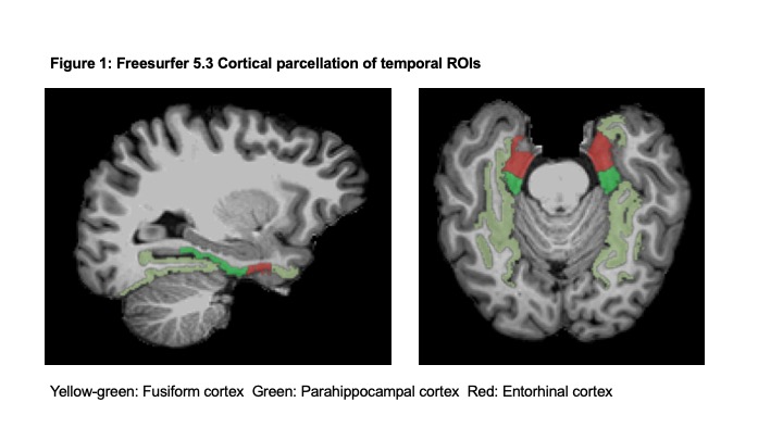
Cortical parcellation of medial temporal cortex using Freesurfer V5.3 atlas indicating the fusiform cortex (yellow-green), the parahippocampal cortex (green), and the entorhinal cortex (red).
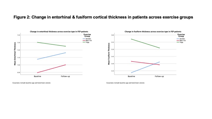
Mean entorhinal cortical thickness and fusiform cortical thickness before and after 12 week exercise intervention for the aerobic (blue), yoga (green), and the waitlist (red) interventions, controlling for age and total brain volume.
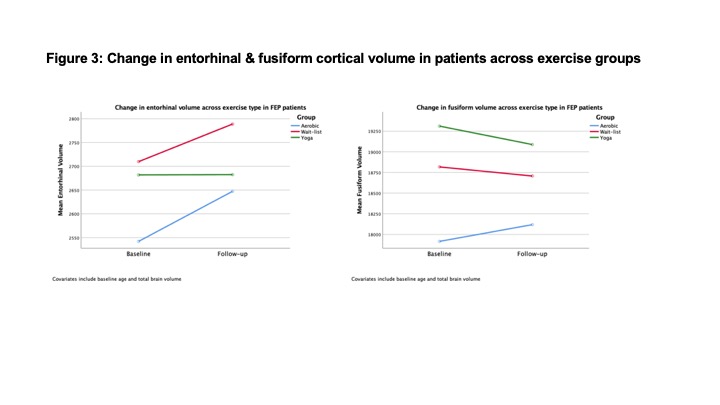
Mean entorhinal cortical volume and fusiform cortical volume before and after 12 week exercise intervention for the aerobic (blue), yoga (green), and the waitlist (red) interventions, controlling for age and total brain volume.
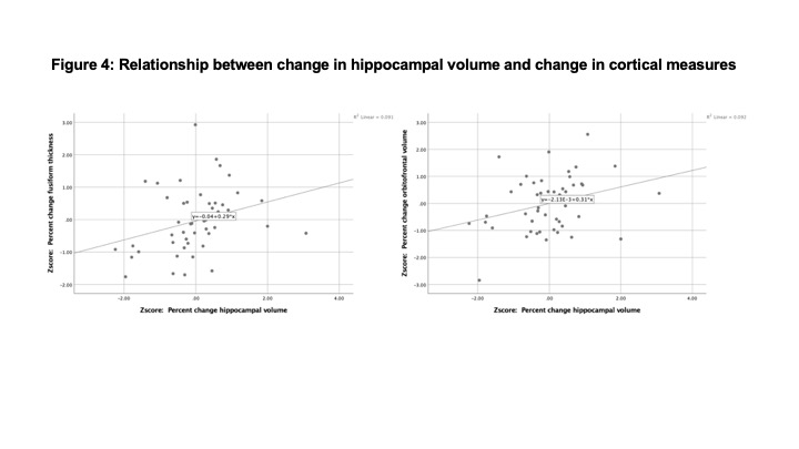
Relationship between percent change in hippocampal volume with fusiform cortical thickness and orbitofrontal cortical volume in early psychosis patients, controlling for age and total brain volume. All data expressed as z-scores.
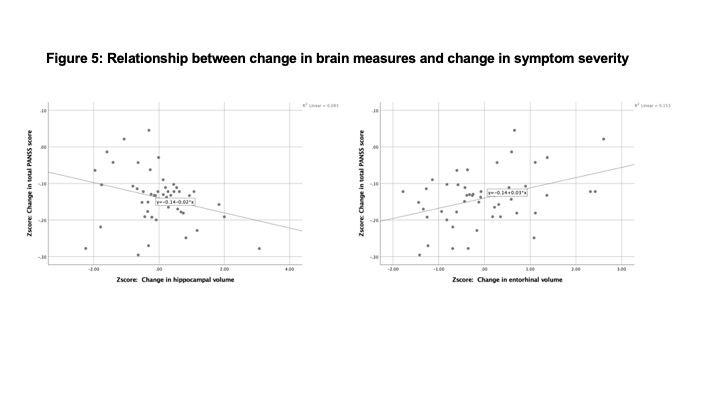
Relationship between percent change in total PANSS (Positive and Negative Syndrome Scale) score with fusiform cortical thickness and orbitofrontal cortical volume in early psychosis patients, controlling for age and total brain volume. All data expressed as z-scores.