2759
Alterations of structural anatomy and functional connectivity regarding hippocampus in obsessive–compulsive disorder1Huaxi MR Research Centre(HMRRC), Department of Radiology, West China Hospital of Sichuan University, Chengdu, China, 2Mental Health Center, Department of Psychiatry, West China Hospital of Sichuan University, Chengdu, China
Synopsis
Besides the classical cortico-striato-thalamo-cortical circuits, the hippocampus has received increasing attention in the psychopathology of obsessive-compulsive disorder (OCD). We aimed to investigate the abnormalities of structural anatomy and functional connectivity (FC) regarding hippocampus in a relatively large sample of unmedicated OCD patients and explore the effects of onset age on these neural correlates. Our findings (i) identified significant volumetric reductions of right hippocampus in OCD; (ii) revealed abnormal cortico-hippocampal connectivity in the prefrontal-limbic networks of OCD and (iii) indicated distinct patterns of cerebral-hippocampal connectivity alterations in early-onset and late-onset OCD, which highlighted the potential importance of neurodevelopmental changes in OCD.
INTRODUCTION
Obsessive-compulsive disorder (OCD) is a neurodevelopmental disorder that affects 1%–3% of the population [1]. 30%-50% of adults with OCD have an onset in childhood rather than in adulthood [2]. Disease models of OCD propose that abnormalities in the cortico-striato-thalamo-cortical circuits are paramount to the psychopathology of OCD [3]. Recent multisite mega-analysis of structural neuroimaging studies [4, 5] and task-based fMRI research [6] also implicate the involvement of mesolimbic regions, especially the hippocampus, in pediatric and adult OCD. However, the functional connectivity (FC) between the hippocampus and the cerebral cortex remained elusive. Thus, we aim to perform seed-based FC analyses to examine abnormalities of cortico-hippocampal connectivity in OCD patients based on resting-state fMRI (RS-fMRI) and evaluate the correlations between cortico-hippocampal FC and clinical features in OCD. Besides, we calculated the total hippocampal volume to explore the associations between structural anatomy and functional connectivity of hippocampus in OCD. Considering the onset age (cut-off age: 20 years) might be a potential marker for the subtyping of OCD [7], we also conducted subgroup analyses of cortico-hippocampal FC for early-onset (<20 years) and late-onset (≥20 years) OCD patients in order to investigate the neurodevelopmental differences between these two populations.METHODS
A total of 88 medication-free OCD patients (45 early-onset OCD patients and 43 late-onset OCD patients) and 88 age, sex and handedness well matched healthy control subjects (HCS) were recruited in current study. The diagnoses of OCD patients were determined by using the structured clinical interview patient edition according to DSM-IV. Clinical symptoms were evaluated using the Yale-Brown Obsessive-Compulsive Scale (Y-BOCS). High-resolution T1-weighted images and RS-fMRI data of all the participants were acquired in a 3.0 T scanner. An automated segmentation pipeline implemented in FreeSurfer software (Version.6.0) (http://surfer.nmr.mgh.harvard.edu/) [8] was used to objectively measure total hippocampal volumes bilaterally. The preprocessing of RS-fMRI was performed using DPABI software (http://www.restfmri.net) [9]. Afterwards, we selected bilateral hippocampuses as regions of interest (ROI) to conduct the seed-based FC analyses for investigating the differences of cortico-hippocampal connectivity in OCD patients compared with HCS using the REST software package (http://resting-fmri.sourceforge.net) [10]. The statistic analyses of main effect (88 OCD patients vs. 88 HCS) and subgroup comparisons (early-onset subgroup: 45 OCD vs. 45 HCS; late-onset subgroup: 43 OCD vs. 43 HCS) regarding cortico-hippocampal FC alterations were performed using the voxel-based two-sample t-test in REST software and the map thresholds were set at P < 0.05 with AlphaSim correction. Pearson correlation analyses were conducted to identify the association between these functional neural correlates and clinical measurements.RESULTS
The structural imaging analyses identified significant decreased volume of right hippocampus in OCD patients compared with HCS (P < 0.05) and this finding remained stable in the following two subgroup analyses (early-onset OCD & late-onset OCD) (Fig 1. box plots). The seed based FC analyses revealed that OCD patients exhibited significantly reduced hippocampus related connectivity in bilateral medial prefrontal cortex (mPFC) and subgenual anterior cingulate cortex (sgACC) as well as left orbit frontal cortex (OFC) relative to HCS and these cortico-hippocampal connectivity reductions were more distributed when the seed ROI was placed in the right hippocampus, which showed volumetric reductions (Fig 1. A). The OFC-hippocampal connectivity was negatively correlated with the illness duration (r = -0.213; P = 0.047) (Fig 1. A orange scatter plot). In the subgroup analyses, early-onset OCD patients exhibited significantly increased FC between the right hippocampus and left superior temporal gyrus and decreased hippocampus related connectivity in bilateral mPFC and left inferior parietal gyrus (IPG) (Fig 1. B); in contrast, the late-onset OCD patients showed reduced hippocampus related connectivity mainly in sgACC and OFC bilaterally (Fig 1. C). Additionally, correlation analysis showed the IPG-hippocampal connectivity was negatively correlated with Y-BOCS total score (r = -0.316; P = 0.034) in the early-onset group (Fig 1. B green scatter plot) while OFC-hippocampal connectivity was negatively correlated with age (r = -0.307; P = 0.045) in the late-onset group (Fig 1. C yellow scatter plot).DISCUSSION & CONCLUSION
The current study provided a thorough profile for structural anatomy alterations and functional connectivity abnormalities of hippocampus in OCD. The altered cortico-hippocampal connectivity in the medial prefrontal and limbic networks, which correlated with illness duration, pointed to a connectivity-based pathophysiologic process in OCD. Furthermore, early-onset OCD is associated with disrupted hippocampus related FC mainly in mPFC and parieto-temporal regions, while late-onset OCD is associated with aberrant hippocampus related FC mainly in OFC and sgACC. These findings indicated distinct patterns of cortico-hippocampal connectivity abnormalities in early-onset and late-onset OCD, which highlighted the potential importance of neurodevelopmental alterations in OCD.Acknowledgements
This study was supported by the National Natural Science Foundation (Grant No. 81671669, 81621003, 81761128023 and 81820108018) and Programme for Changjiang Scholars and Innovative Research Team in University (PCSIRT, Grant No. IRT16R52) of China. Dr. Qiyong Gong would like to acknowledge his Visiting Adjunct Professor appointment in the Department of Psychiatry at the Yale School of Medicine, Yale University, USA. The authors reported no biomedical financial interests or potential conflicts of interest.References
1. Ruscio, A.M., et al., The epidemiology of obsessive-compulsive disorder in the National Comorbidity Survey Replication. Mol Psychiatry, 2010. 15(1): p. 53-63.
2. Pauls, D.L., et al., Obsessive-compulsive disorder: an integrative genetic and neurobiological perspective. Nat Rev Neurosci, 2014. 15(6): p. 410-24.
3. Dougherty, D.D., et al., Neuroscientifically Informed Formulation and Treatment Planning for Patients With Obsessive-Compulsive Disorder: A Review. JAMA Psychiatry, 2018. 75(10): p. 1081-1087.
4. Boedhoe, P.S., et al., Distinct Subcortical Volume Alterations in Pediatric and Adult OCD: A Worldwide Meta- and Mega-Analysis. Am J Psychiatry, 2017. 174(1): p. 60-69.
5. Fouche, J.P., et al., Cortical thickness in obsessive-compulsive disorder: multisite mega-analysis of 780 brain scans from six centres. Br J Psychiatry, 2017. 210(1): p. 67-74.
6. Marsh, R., et al., Reward-based spatial learning in unmedicated adults with obsessive-compulsive disorder. Am J Psychiatry, 2015. 172(4): p. 383-92.
7. Anholt, G.E., et al., Age of onset in obsessive-compulsive disorder: admixture analysis with a large sample. Psychol Med, 2014. 44(1): p. 185-94.
8. Fischl, B., et al., Whole brain segmentation: automated labeling of neuroanatomical structures in the human brain. Neuron, 2002. 33(3): p. 341-55.
9. Yan, C.G., et al., DPABI: Data Processing & Analysis for (Resting-State) Brain Imaging. Neuroinformatics, 2016. 14(3): p. 339-51.
10. Song, X.W., et al., REST: a toolkit for resting-state functional magnetic resonance imaging data processing. PLoS One, 2011. 6(9): p. e25031.
Figures
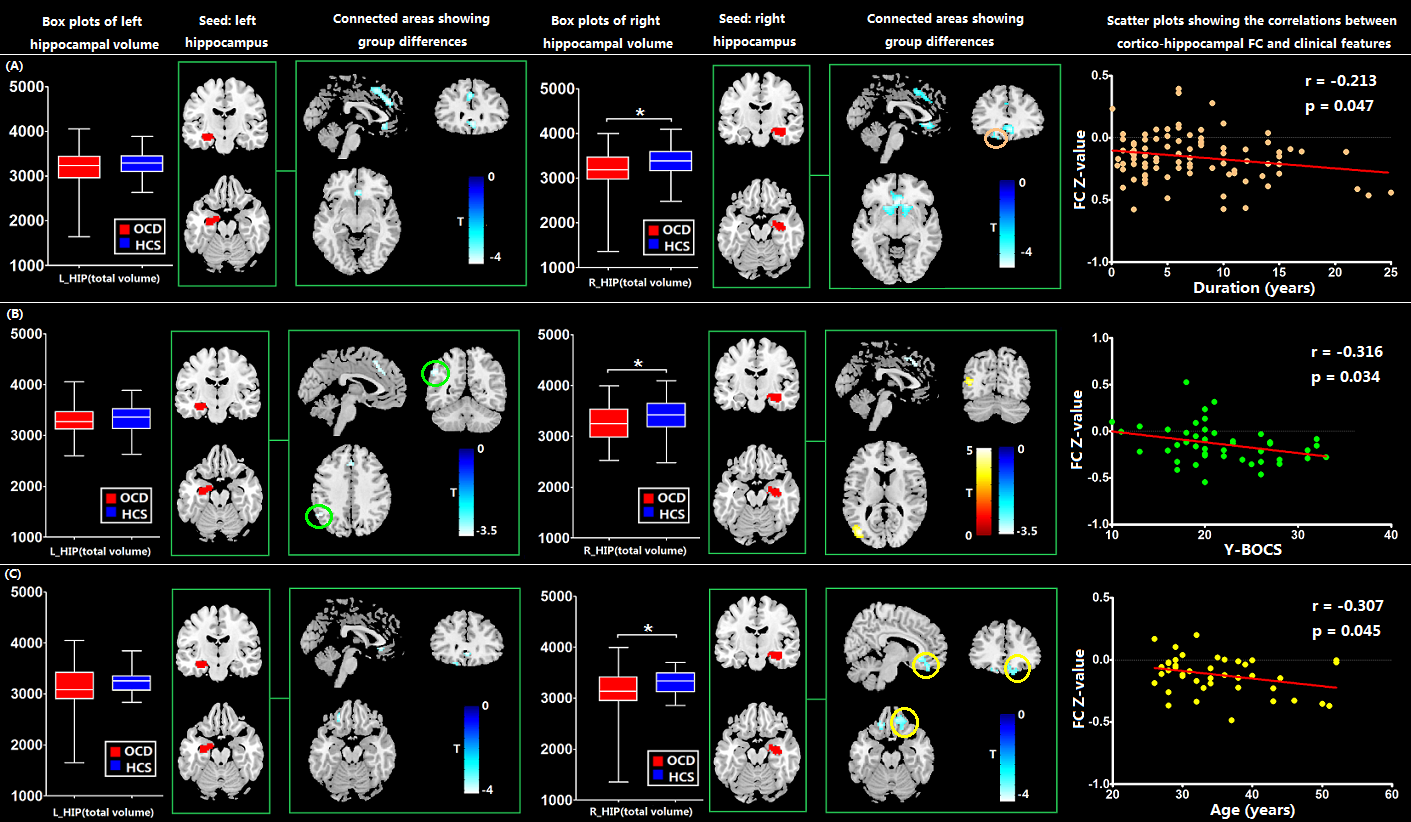
Fig 1: The statistical analyses regarding the hippocampal volumetric alterations and cortical-hippocampal FC abnormalities between OCD patients and HCS as well as the findings of correlation analysis between the averaged eigenvalues of FC and the clinical features. (A) main effect: 88 OCD patients vs. 88 HCS; (B) subgroup analysis of early-onset group: 45 early-onset OCD patients vs. 45 HCS; (C) subgroup analysis of late-onset group: 43 late-onset OCD patients vs. 43 HCS. FC increases are indicated in warm colors while FC reductions are indicated in cool colors.
Abbreviations: HCS, healthy control subjects; OCD, obsessive-compulsive disorder; FC, functional connectivity