2754
Gray matter network changes with aging and duration in a large group of never-treated patients with schizophrenia.1Department of Radiology, West China Hospital, Sichuan University., Chengdu, China, 2West China Hospital, Sichuan University., Chengdu, China
Synopsis
A large group of never-treated schizophrenia patient was enrolled to investigate the pattern of network changes with aging and illness duration. Cortical thickness preprocessed by FreeSurfer, and correlation matrix was constructed by correlating the cortical thickness of every pair of regions. Compared to healthy controls, all patient subgroups stratified along age and illness duration showed common changes while distinct changes, mainly involved DMN and CN. The alternations within DMN and CN may represent trait-related structural network changes in schizophrenia, while distinct changes may represent illness progression with more-wide spread brain abnormalities.
INTRODUCTION
Abnormal structural brain networks are believed to be an important neuropathology of serious mental illness. The knowing of brain network changes with aging could advance our understanding of these network disturbances and potential pathophysiology of schizophrenia. The primary hypothesis about progressivity of schizophrenia has been posed by Kraepelin in Dementia Praecox and Paraphrenia (1919), and accumulated papers were published in rapid succession providing the progressive structure changes of schizophrenia,1-3 although with inconsistency. In the present study, we enrolled a large group of never-treated schizophrenia patient and focused on nodal property changes to investigate the pattern of network changes with aging and illness duration. And more specifically, whether there exists progressivity in gray matter network changes in schizophrenia along aging and duration.METHODS:
In this program, we enrolled 152 never-treated schizophrenia patients, whose illness initiated after 15 years old, and 201 healthy controls, all patients were stratified respectively into 4 subgroups A1-A4 by age (16-24, 25-34, 35-44,>45), and into D1-D4 by illness duration (<12 months, 12-60 months, more than 60 months), then matched controls for each subgroup were selected from 201 healthy subjects. High resolution T1 images of brain were obtained with a 3.0 T MR scanner (GE), and preprocessed by FreeSurfer. The gray matter network matrices were constructed by correlating the cortical thickness of every pair of regions within AAL brain segmentations across individuals.4 The network properties including nodal centrality and efficiency were then calculated.RESULTS:
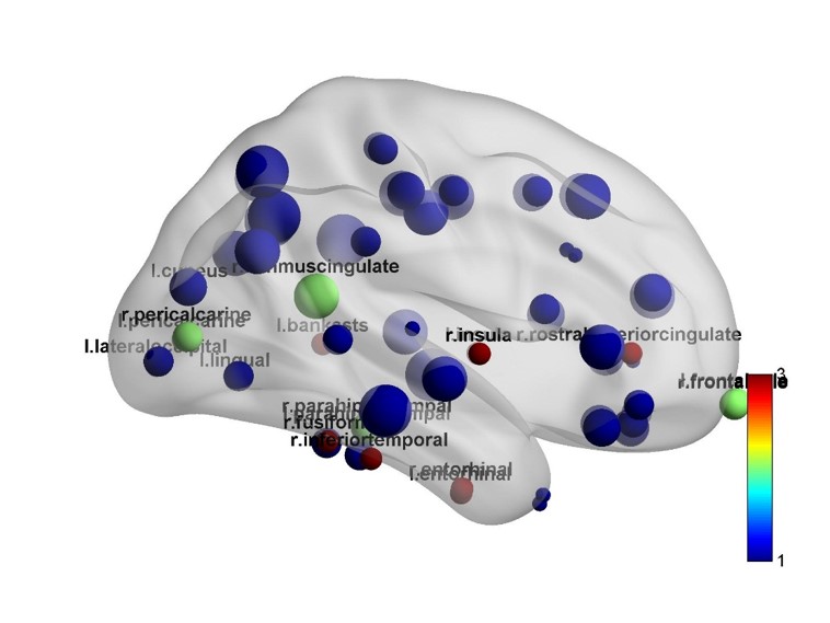
Compared to healthy controls, all patient subgroups stratified along age showed some common network property changes while some distinct changes. The common changes along aging were nodal centrality loss at regions mainly within default mode network (DMN), control network (CN). More importantly, the network deteriorates with aging and more nodal centrality decreases at isthmus cingulate, right caudal anterior cingulate were showed at subgroups A2-A4, and more abnormality of left precuneus showed at A3-A4. All patient subgroups stratified along duration showed common network property changes at core area of DMN and CN, right isthmus cingulate, left caudal and rostral anterior cingulate, besides that, they also showed common changes at bilateral inferior temporal and entorhinal, left para hippocampal and pericalcarine, and right medial and lateral orbitofrontal. Except that, more network centrality loss at regions of DMN, CN and visual network showed at subgroup D2-D3.DISCUSSION:
Age and illness duration are closely related variables, each stratification method has different lay point. The changes of DMN in schizophrenia had been repeatedly demonstrated by a large quantity of papers. 5-7 Functional MRI research indicates that DMN and CN have interaction in schizophrenia.8 These common changes showed respectively in all subgroups of age and illness duration suggest there do exist stable gray matter network changes in DMN and CN, which might contribute to the interaction evidence. Subgroups of either grouping strategy showed nodal properties loss in more brain regions at later aging and illness duration, which would indicate the deterioration of illness, even in a relatively less significant level. As we can see, results of age subgroups and illness duration subgroups have overlaps and inconsistency.CONCLUSION:
The alternations of topological properties within DMN and CN may represent trait-related structural network changes in schizophrenia, while the changes at later aging and illness duration may represent illness progression with more-wide spread brain abnormalities.Acknowledgements
Address correspondence to Dr. Lui (lusuwcums@tom.com).
Supported by the National Natural Science Foundation (grants 81222018,81371527, and 81401396)
References
1. Xiao Y, Sun H, Shi S, et al. White Matter Abnormalities in Never-Treated Patients With Long-Term Schizophrenia. The American journal of psychiatry 2018:appiajp201817121402.
2. Shah C, Liu J, Lv P, et al. Age Related Changes in Topological Properties of Brain Functional Network and Structural Connectivity. Frontiers in neuroscience 2018;12:318.
3. Cropley VL, Klauser P, Lenroot RK, et al. Accelerated Gray and White Matter Deterioration With Age in Schizophrenia. The American journal of psychiatry 2017;174:286-95.
4. He Y, Chen Z, Evans A. Structural insights into aberrant topological patterns of large-scale cortical networks in Alzheimer's disease. J Neurosci 2008;28:4756-66.
5. Hu ML, Zong XF, Mann JJ, et al. A Review of the Functional and Anatomical Default Mode Network in Schizophrenia. Neuroscience bulletin 2017;33:73-84.
6. Pankow A, Deserno L, Walter M, et al. Reduced default mode network connectivity in schizophrenia patients. Schizophrenia research 2015;165:90-3.
7. Guo W, Su Q, Yao D, et al. Decreased regional activity of default-mode network in unaffected siblings of schizophrenia patients at rest. European neuropsychopharmacology : the journal of the European College of Neuropsychopharmacology 2014;24:545-52.
8. Manoliu A, Riedl V, Zherdin A, et al. Aberrant dependence of default mode/central executive network interactions on anterior insular salience network activity in schizophrenia. Schizophrenia bulletin 2014;40:428-37.
Figures