2750
Contribution and Interaction of Brain Structure and Function in Treatment Response Prediction of First-episode Drug-naïve Schizophrenia1Radiology, West China Hospital, Sichuan University, Chengdu, China, 2Institution of Psychology, CAS, Beijing, China
Synopsis
Magnetic Resonance Imaging (MRI) including structural and functional neuroimaging has been applied extensively to examine response to antipsychotic treatment, however, questions remain regarding the interaction between these measurements and their unique role. Our study provided a comprehensive examination of interaction and contribution for voxel-wise measurements related with treatment response. We found that brain functional measurements in certain brain regions have advantages in predicting treatment response. Furthermore, the functional activities were different between short- and long-term treatment of antipsychotic drugs. These findings revealed that functional changes were more sensitive to the antipsychotic treatment and could be promising biomarkers in treatment prediction.
Introduction
Brain structural and functional neuroimaging biomarkers have been widely used to track and predict treatment outcomes to antipsychotics in first-episode schizophrenia 1. However, it is difficult to establish their own weights and contributions in the outcome prediction. The present study aimed to investigate the interaction between brain structural and functional indices and their respective roles in the prediction of treatment response to antipsychotics in schizophrenia.Methods
A total of 83 first-episode drug-naïve patients with schizophrenia underwent three dimensional T1 and resting-state functional MR (R-fMRI) scans and among them, 42 patients completed 6-weeks of follow-up and 64 patients completed one-year of follow-up, respectively. Symptom severity was assessed by Positive and Negative Symptom Scale (PANSS) and treatment response was quantified by the percentage changes in PANSS total scores 2. The method applied in the present study was published in our previous study 3. Briefly, we selected one of two structural parameters including gray matter volume (GMV) and gray matter density (GMD) and one of five functional measurements including amplitude of low frequency fluctuations (ALFF), fractional amplitude of low frequency fluctuations (fALFF), regional homogeneity (ReHo), voxel-mirrored homotopic connectivity (VMHC) and network degree centrality (DC) to calculate a voxel-wise interaction index. Then, a general linear model was constructed to explore the relationship between the structural-functional interaction indices across all brain voxels for each participant and treatment response. Finally, to further address which MR measure is the most important predictor of the treatment response, a variation decomposition was conducted in the brain masks that show a significant relationship between treatment response and interaction indices. Gaussian random field correction was used for multiple comparison correction 4,5. Voxel-wise p<0.001 and cluster-wise p<0.05 were statically significant.Results
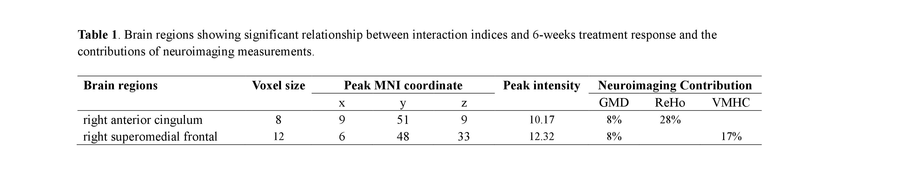
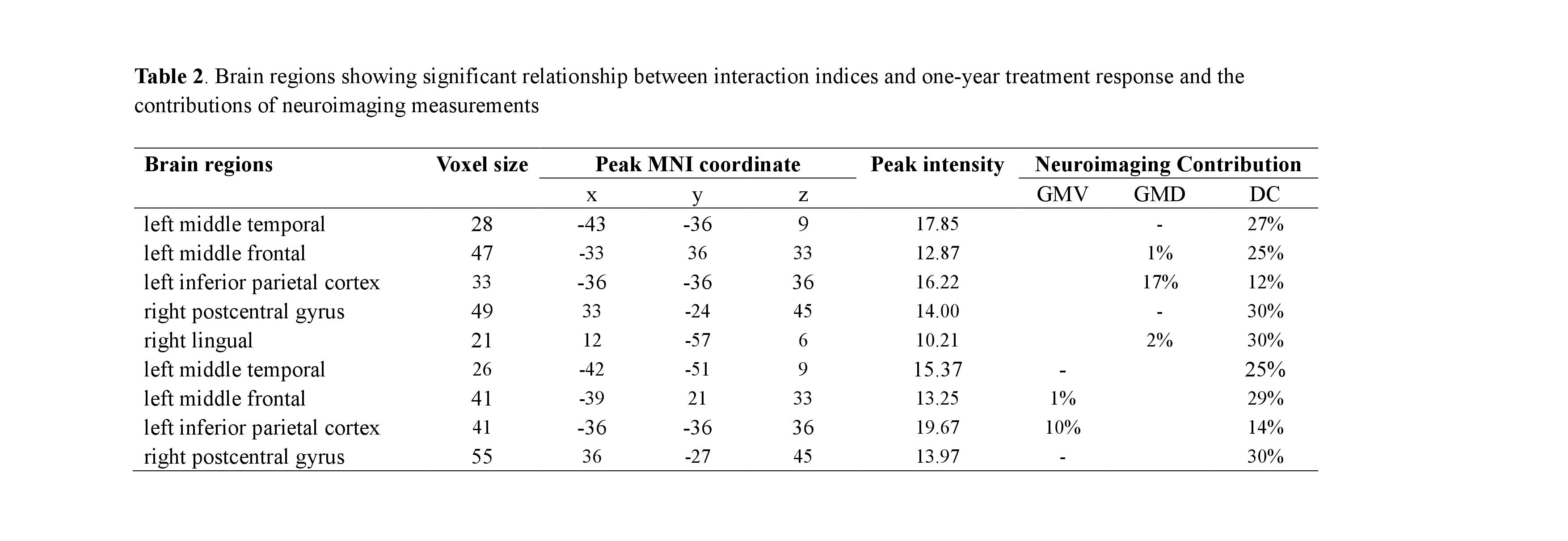
Voxel-wise interaction between structural and functional measurements was related to treatment response. The interaction indices derived from GMD and ReHo in the right anterior cingulum and from GMD and VMHC in the right superomedial frontal cortex were correlated with six-weeks treatment response. Additionally, the functional measurements have greater contribution to the response than structural measurements (see Figure 1). The interaction indices derived from GMV and DC in the left middle frontal cortex, left inferior parietal cortex, left middle temporal lobe, right lingual and right postcentral gyrus and from GMD and DC in the brain regions as similar as the regions before except the right lingual lobe were correlated with one-year treatment response. Furthermore, DC was more closely related with treatment response (see Figure 2).Discussion
The present study found that functional measurements had a closer relationship with treatment response than structural parameters, suggesting that functional measurements are better biomarkers for outcome prediction within one-year treatment. Notably, the present study showed that the functional measurements related to treatment response at 6 weeks and one year were different. The measurements associated with short-term response mainly reflect the local activities (i.e., ReHo), which was consistent with previous studies 6,7. However, the parameter associated with long-term treatment reflects global connectivity (i.e., DC). We speculated that antipsychotic drugs have local effects in acute treatment trial and have extensive impact on brain networks after long-term treatment.Conclusion
The present study provided evidence that brain functional activities in certain brain regions take advantages in predicting treatment response. Furthermore, the study found that changes of functional activities were associated with treatment duration, suggesting that functional changes are more sensitive to the antipsychotic treatment and could be promising biomarkers in treatment prediction.Acknowledgements
This study was supported by the National Natural Science Foundation of China (GrantsNos. 81371527, 81671664, and 81621003). Dr. Lui would also like to acknowledge the support from Chang Jiang Scholars (Award No. Q2015154) of China, and the National Program for Support of Top-notch Young Professionals (National Program for Special Support of Eminent Professionals, Organization Department of the Communist Party of China Central Committee, Award No. W02070140).References
1. Tarcijonas, G. & Sarpal, D. K. Neuroimaging markers of antipsychotic treatment response in schizophrenia: An overview of magnetic resonance imaging studies. Neurobiol. Dis. [Epub], (2018). 2. Cao, B. et al. Treatment response prediction and individualized identification of first-episode drug-naïve schizophrenia using brain functional connectivity. Mol. Psychiatry [Epub], (2018). 3. Yan, C. G., Yang, Z., Colcombe, S. J., Zuo, X. N. & Milham, M. P. Concordance among indices of intrinsic brain function: Insights from inter-individual variation and temporal dynamics. Sci. Bull. 62, 1572–1584 (2017). 4. Nichols, T. & Hayasaka, S. Controlling the familywise error rate in functional neuroimaging: a comparative review. Stat. Methods Med. Res. 12, 419–446 (2003). 5. Friston, K. J., Worsley, K. J., Frackowiak, R. S., Mazziotta, J. C. & Evans, A. C. Assessing the significance of focal activations using their spatial extent. Hum. Brain Mapp. 1, 210–220 (1994). 6. Gao, S. et al. Distinguishing Between Treatment-Resistant and Non-Treatment-Resistant Schizophrenia Using Regional Homogeneity. Front. Psychiatry 9, 1–9 (2018). 7. Lui, S. et al. Short-term effects of antipsychotic treatment on cerebral function in drug-naive first-episode schizophrenia revealed by ‘resting state’ functional magnetic resonance imaging. Arch. Gen. Psychiatry 67, 783–792 (2010).Figures