2749
Myelin-associated clinical and physical correlates in a cohort of chronic schizophrenia patients.1Radiology, University of British Columbia, Vancouver, BC, Canada, 2University of British Columbia, Vancouver, BC, Canada, 3Psychiatry, University of British Columbia, Vancouver, BC, Canada, 4Psychology, Simon Fraser University, Burnaby, BC, Canada, 5Pathology & Laboratory Medicine, University of British Columbia, Vancouver, BC, Canada, 6Physics & Astronomy, University of British Columbia, Vancouver, BC, Canada, 7International Collaboration on Repair Discoveries (ICORD), University Of British Columbia, Vancouver, BC, Canada, 8Kinesiology, University of British Columbia, Vancouver, BC, Canada
Synopsis
Aberrant myelination and tandem cardiovascular deficits may contribute to emergence of the schizophrenias. To explore this hypothesis, a pilot study of Myelin Water Fraction (MWF), V02max capacity, and symptom severity was done in 15 chronic schizophrenia/schizoaffective patients. MWF was positively correlated with age in some, but not all, fronto-medial and fronto-temporal regions, 2. V02max was positively correlated with MWF the superior longitudinal fasciculus, the genu, and the forceps minor, and 3. Social functioning was positively correlated to MWF in the forceps major. These data indicate the presence of relationships between MWF measures, social functioning and cardiovascular capacity in schizophrenia.
Background Rationale/Purpose
Myelin, which mediates normal signal conductance in the brain, experiences cyclical bursts of growth throughout the lifespan and is thought to be influenced by cardiovascular health1. Aberrant myelination is thought to contribute to the emergence of mental disorders, including schizophrenia. However, structure-function relationships in schizophrenia have long been controversial with inconsistent findings. The complexity of such psychotic disorders is more suggestive of diffuse dysconnectivity between key brain areas leading to the emergence of clinical symptoms2. Adverse interactions with poor underlying cardiovascular health may further exacerbate poor general functioning in patients3. We previously demonstrated that dysmyelination may be a key contributor to dysconnectivity in schizophrenia4,5 and myelin deficits in frontal and temporal regions are believed to have damaging functional outcomes for patients. In this pilot investigation our objective was to explore the relationships between cardiovascular capacity, social functioning and MR-measured myelin in a cohort of chronic schizophrenia patients.Methods
Data acquisition: Imaging was performed on a Philips 3T Achieva scanner in 9 chronic DSM-V schizoaffective and 6 schizophrenia patients (mean age 31.1yrs, age range: 23.0-40.5yrs, 6 F, 9 M) with a SENSE-Head 8 coil. Data collected included myelin water imaging (3D Gradient Spin Echo (GRASE), 32-echo, TR/TE = 1000/10ms, 40 contiguous transverse slices, slice thickness = 3 mm, FOV = 230mm2, reconstruction resolution = 0.96x0.95x2.5mm, flip angle = 90°) and a 3DT1 volumetric scan (TR/TE = 6.6/3.0ms, 155 sagittal contiguous slices, slice thickness = 1 mm, FOV = 240mm2, reconstruction resolution = 1x1x1mm, flip angle = 8°).
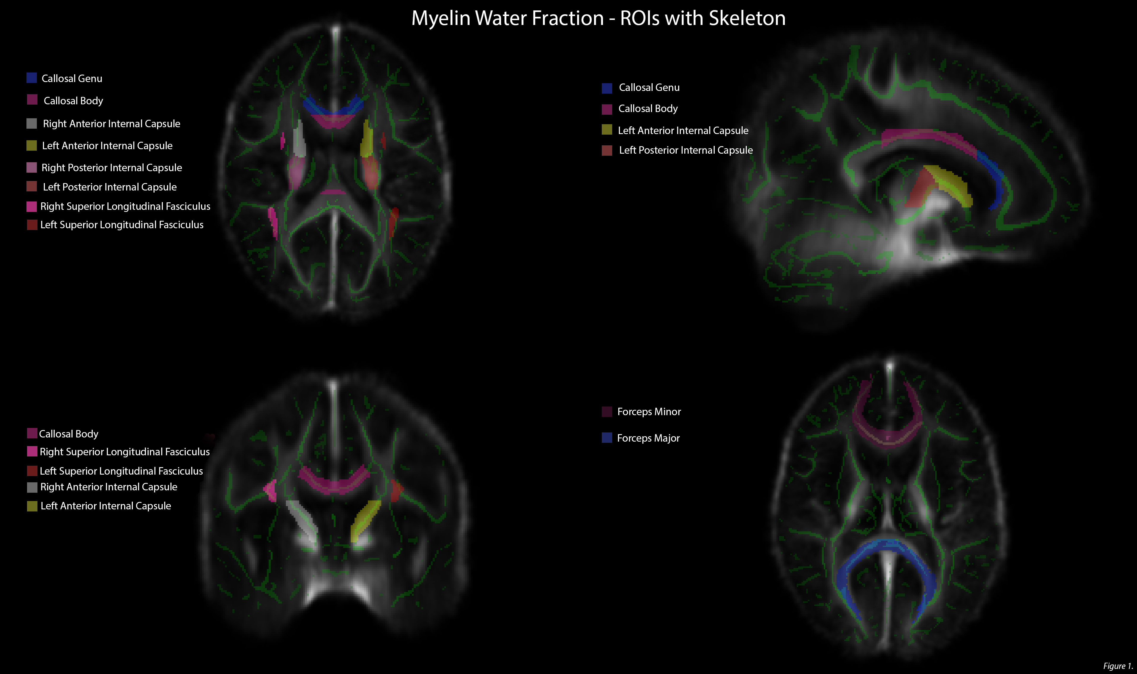
Data analysis: All scans were visually inspected by a trained neuroradiologist for abnormalities. GRASE data was mapped to 3D volumetric space in FSL 5.0.16. White matter regions of interest were extracted using the JHU atlas7,8,9. Voxel-wise T2 distributions were calculated from the GRASE data using a modified Extended Phase Graph algorithm combined with regularized non-negative least squares and flip angle optimization10,11. MWF was defined as the fraction of signal with T2<40ms12,13. Mean MWF was determined for each ROI. Left and right hemisphere MWFs were examined separated and as pooled means.
Statistical analysis: Exploratory linear correlation models were used to probe MWF relationships to clinician rated symptom severity, cardiovascular capacity (V02max – an index of maximal oxygen uptake)14. Cognitive and general functioning measures were taken using the Symbol Digits Modality Test (SMDT)15, Social and Occupational Functioning Assessment Scale16, and the Calgary Depression Scale17. Bonferroni adjustment was not applied to the current results, given the exploratory nature of this pilot study.
Results
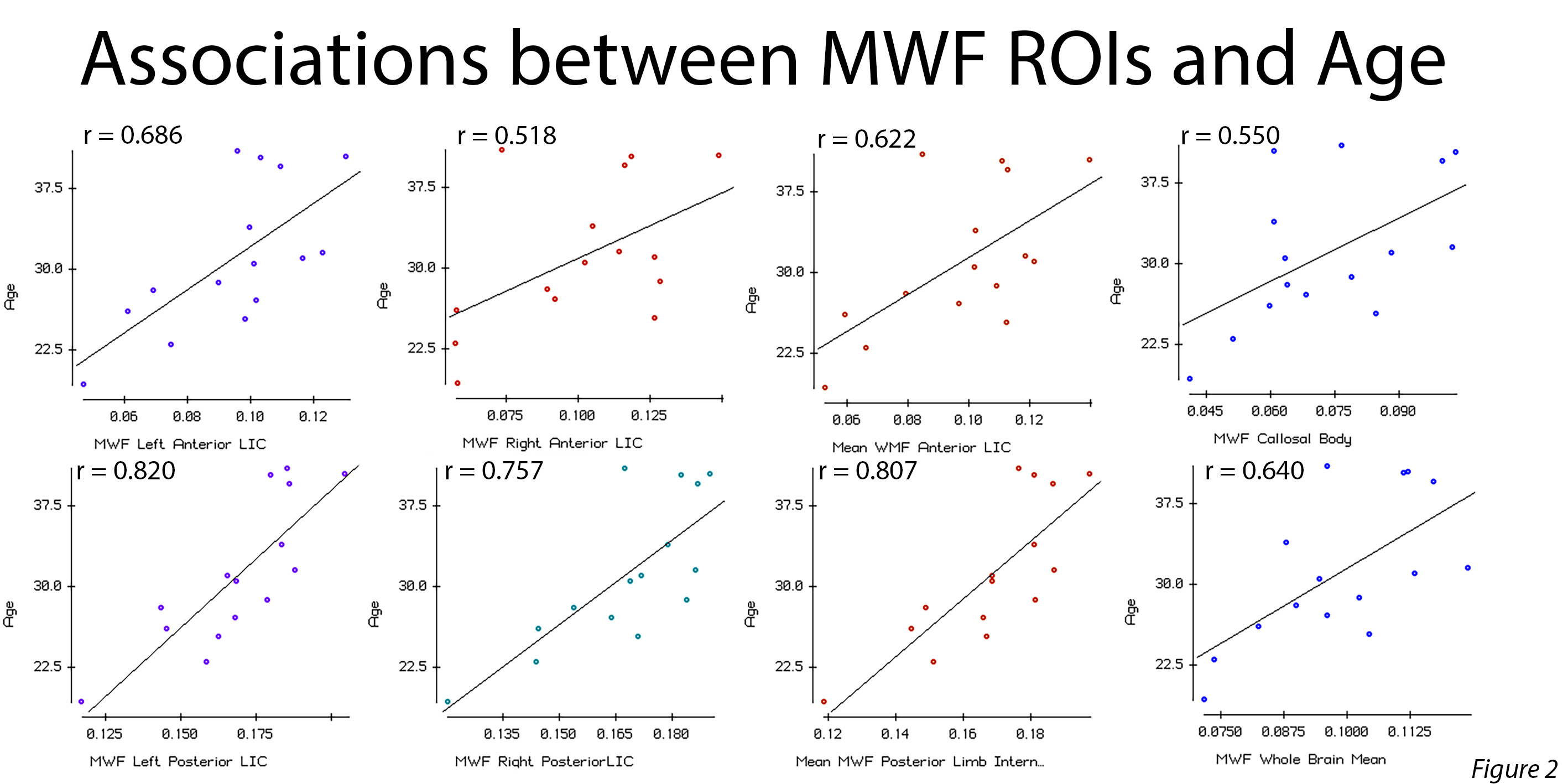
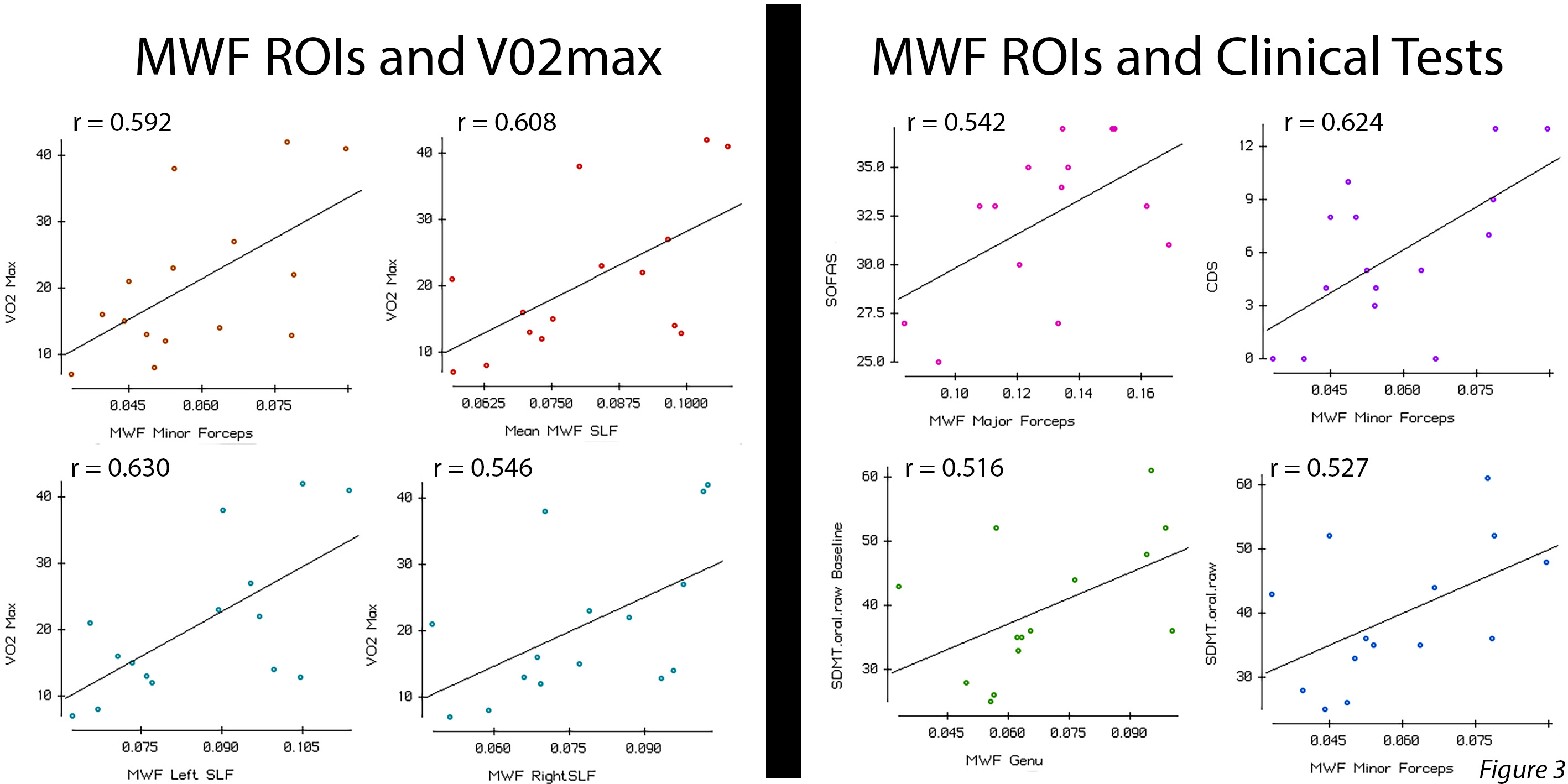
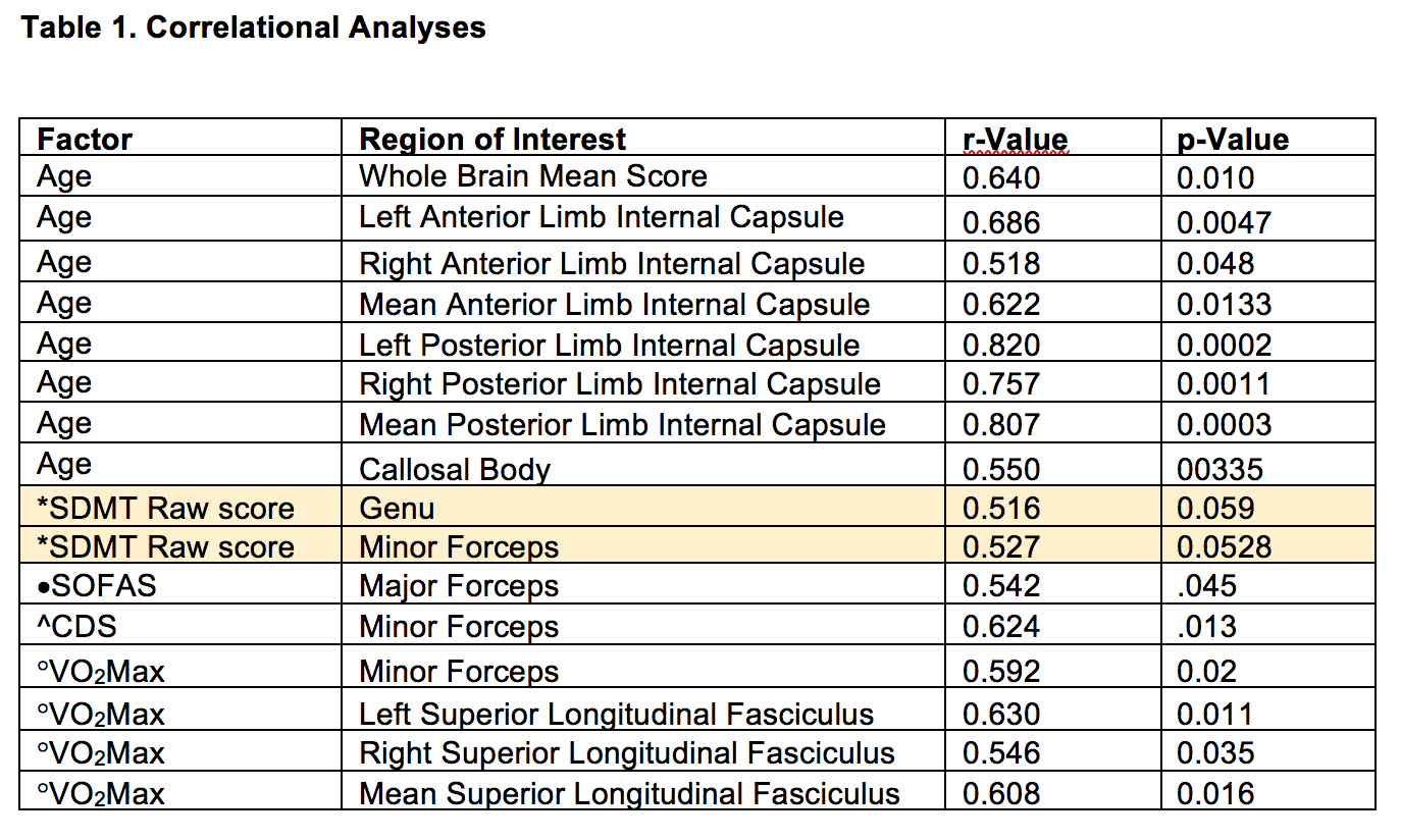
Strong positive relationships between age and bilateral MWFs in the anterior and posterior limbs were observed (Figure 1), particularly on the left (Anterior Limb: r = 0.686, p. = 0.0047, Posterior Limb r = 0.820, p. = 0.0003). No significant age relationships to any other white matter region were seen. MWF was not associated with symptom severity in any region, as indicated by the PANSS (all p-values > 0.10) nor was it associated with antipsychotic dose (all p-values > .20). No relationships between MWF in region and IQ or educational attainment were observed in this group (all p-values > 0.30). A strong trend for positive associations between measures of cognitive processing speed and MWF in the genu and the forceps minor was observed (p-values = 0.059 and 0.0523, respectively). A significant relationship between social functioning and MWF in the forceps major was seen. Cardiovascular capacity was positively associated with MWF scores in the genu, the forceps minor, and bilaterally with the superior longitudinal fasciculi. MWF scores were not associated with over all brain volume (all p-values >.05). See MWF Images are shown in Figure 1 with graph for the data in Figures 2 and 3. A summary of the correlational analyses can be found in Table 1.Conclusions
As expected, we observed relationships between age and MWF in deep white matter paths. This was not observed for frontal or temporal areas, in contrast to our previous findings in younger early psychosis patients and healthy volunteers5. Strong trends were observed in SDMT scores and MWF in frontal areas. Given the small sample size, this study may be under-powered to detect more moderate effects. We were able to demonstrate relationships between cardiovascular capacity and MWF in frontal and fronto-temporal paths, demonstrating the potential benefits to white matter integrity of better cardiovascular health. Further investigations will be required to determine how much underlying cardiovascular capacity affects myelin integrity and cognition.Acknowledgements
We would like to thank the Canadian Institutes of Health Research, the Mind Foundation of British Columbia, and the BC Provincial Health Services Authority for grant funding. In addition we want to acknowledge all the participants who took part in this study, study volunteers, and the MRI technicians at the UBC MRI Research Centre who helped make this research possible.
References
1. Yeatman JD, Wandell BA, Mezer AA. Lifespan maturation and degeneration of human brain white matter. Nat Commun. 2014;5:4932.
2. Friston K, Brown HR, Siemerkus J, Stephan KE. The dysconnection hypothesis (2016). Schiz Res. 2016;176:83-93.
3. De Hert M, Detraux J, Psy M, Vancampfort D. The intriguing relationship between coronary heart disease and mental disorders. Dialogues Clin Neurosci. 2018;20:31-40.
4. Flynn SW, Lang DJ, Mackay AL, Goghari VM, Vavasour IM, Whittall KP, Smith GN, Arango V, Mann JJ, Dwork AJ, Falkai P, Honer WG. Abnormalities of myelination in schizophrenia detected in vivo with MRI, and post-mortem with analysis of oligodendrocyte proteins. Molecular Psychiatry. 2003;8:811-820.
5. Lang DJ, Yip E, MacKay AL, Thornton AE, Vila-Rodriguez F, MacEwan GW, Smith GN, Laule C, MacCrae CB, Honer WG. 48 Echo T2 myelin imaging in first-episode schizophrenia: evidence of aberrant myelination. Neuroimage:Clin. 2014;4:408-414.
6. Jenkinson M, Beckmann CF, Behrens TE, Woolrich MW, Smith SM. FSL. NeuroImage, 62:782-90, 2012
7. Mori S, Wakana S, van Zijl PCM, Nagae-Poetscher LM. MRI Atlas of Human White Matter. Elsevier, Amsterdam, The Netherlands (2005)
8. Wakana S, Caprihan A, Panzenboeck MM, Fallon JH, Perry M, Gollub RL, … Mori S. Reproducibility of quantitative tractography methods applied to cerebral white matter. NeuroImage 36:630-644 (2007)
9. Hua K, Zhang J, Wakana S, Jiang H, Li X, Reich, D, … Mori S. Tract probability maps in stereotaxic spaces: analysis of white matter anatomy and tract-specific quantification. NeuroImage, 39(1):336-347 (2008)
10. Prasloski T, Mädler B, Xiang Q-S, et al. Applications of stimulated echo correction to multicomponent T2 analysis. Magn Reson Med 2012; 67: 1803–1814.
11. Whittall KP, Mackay AL, Graeb DA, Nugent RA, Li DK, Paty DW. In vivo measurement of T2 distributions and water contents in normal human brain. Magnetic Resonance in Medicine. 1997;37:34-43.
12. MacKay AL, Whittall KP, Adler J, Li DK, Paty DW, Graeb DA. In vivo visualization of myelin water in brain by magnetic resonance. J Soc Mag Res Med. 1994;31:673-677.
13. Meyers SM, Laule C, Vavasour IM, Kolind SH, Madler B, Tam R, Traboulsee AL, Lee J, Li DK, Mackay AL. Reproducibility of myelin water fraction analysis: a comparison of region of interest and voxel-based analysis methods. Magnetic Resonance Imaging. 2009;27:1096-1130.
14. Warburton DE, Nicol C, Bredin SS. Health benefits of physical activity: the evidence. Can. Med. Assoc. J. 2006;174:801-809.
15. Benedict RH, DeLuca J, Phillips G, LaRocca N, Hudson LD, Ruckick R. Validity of the Symbol Digit Modalities Test as a cognition performance outcome measure for multiple sclerosis. Mult Scler. 2017;23:721-733.
16. Morosini PL, Magliano L, Bramilla L, Ugolini S, Pioli R. Development, reliability and acceptability of a new version of the DSM-IV Social and Occupational Functioning Assessment Scale (SOFAS) to assess routine social functioning. Acta Psychiatr Scand. 2000;10.
17. Addington, D, Addington, J. & Maticka-tyndale, E (1993). Assessing Depression in Schizophrenia: The Calgary Depression Scale. British Journal of Psychiatry, 163(S22), 39–44. https://doi.org/10.1192/S0007125000292581
Figures