2745
Use of NODDI for Microstructural Characterization of Posterior Limb of the Internal Capsule in Subacute and Chronic Stroke Patients1Istituto di Bioimmagini e Fisiologia Molecolare, Consiglio Nazionale delle Ricerche, Segrate, Italy, 2Istituto di Neuroscienze, Consiglio Nazionale delle Ricerche, Milano, Italy, 3Laboratory of Brain Pathology and Pharmacology and Neuro Center, Humanitas Clinical and Research Center, Rozzano, Italy, 4Neuroradiology Unit and Neuro Center, Humanitas Clinical and Research Center, Rozzano, Italy, 5Stroke Unit and Neuro Center, Humanitas Clinical and Research Center, Rozzano, Italy, 6Neurorehabilitation Unit and Neuro Center, Humanitas Clinical and Research Center, Rozzano, Italy
Synopsis
This preliminary work shows that NODDI, supplementing the classical DTI approach, could provide a new insight into the subtle micro-architectural modifications occurring in the posterior limb of the internal capsule of 10 stroke patients in subacute and chronic phase.
Introduction
Stroke is worldwide the second leading cause of death and in western countries the second leading cause of long-term disability. Provide information about corticospinal tract (CST) integrity, and in particular at the level of the Posterior Limb of the Internal Capsule (PLIC), can improve predictions on patients’ motor outcome and the size of CST injury evaluated using DTI can be a prognostic factor that determines motor performance and outcome in stroke patients1. Neurite Orientation Dispersion and Density Imaging (NODDI) is a model-based imaging technique that can reveal microstructural features of brain tissue in several diseases including stroke2. The aim of the paper is to evaluate if NODDI, supplementing the classical DTI approach, could provide a new insight into the subtle micro-architectural modifications occurring in the PLIC, of stroke patients in subacute and chronic phases.Methods
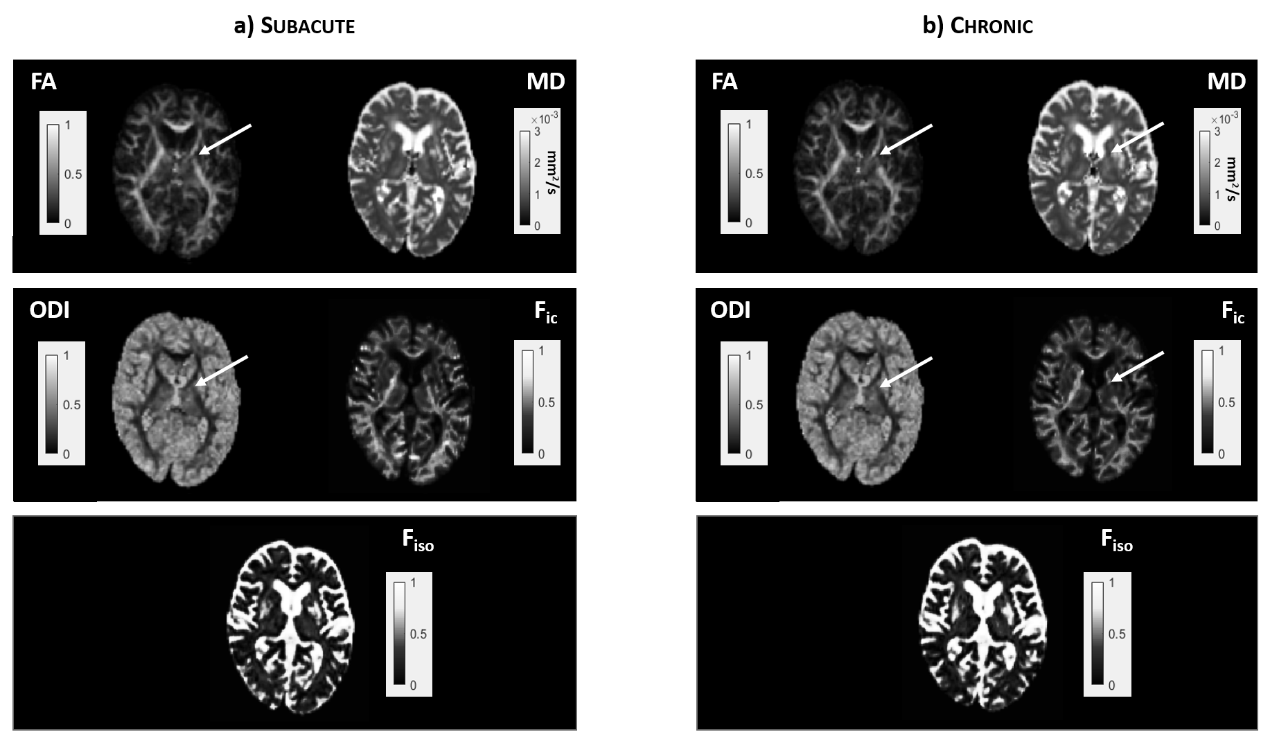
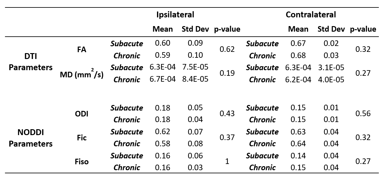
17 subjects (age: 68±11 years) in the subacute phase (14±3 days post-stroke), 10 of whom re-scanned in the chronic phase (231 ± 36 days post-stroke), were enrolled in this study. Images were acquired using a 3T MRI scanner. A two shells EPI protocol was implemented with the following parameters: shell 1- 20 gradient directions; b=700 s/mm2; 3 b=0; shell 2- 64 gradient directions; b=2000 s/mm2; 9 b=0. Diffusion images were preprocessed and DTI maps (Fractional Anisotropy - FA and Mean Diffusivity - MD) were calculated using the software ExploreDTI. NODDI parameters (Orientation Dispersion Index - ODI, Intracellular Volume Fraction – Fic and Isotropic Fraction - Fiso) were estimated using the dedicated Matlab toolbox. The analysis focused on the PLIC. ROIs were manually outlined and checked by an expert neurologist. Statistical analyses were performed by two-sided Wilcoxon signed rank test along with Benjamini-Hochberg correction for multiple comparisons.Results
In the subacute phase, a reduction (-10%; p=0.019) of FA values was correlated with an increase of ODI (+24%; p=0.037), in both the ipsilesional PLIC, suggesting that increased fiber dispersion can be the main structural factor which is altered in this phase. In the chronic phase, a reduction of FA (-13%; p=0.001) and an increase of ODI (+ 21%; p=0.019) persisted in the ipsilesional areas. This was associated with a reduction of Fic (up to -24%; p=0.001) and an increase of MD (up to +8%; p=0.037), suggesting that in the chronic phase fiber reduction, possibly due to nerve degeneration, could play an important role. For further details please see figure 1 and figure 2.Discussion
In subacute stage, the FA alteration was not accompanied by altered MD values in the lesioned hemispheres compared to the contralateral due the pseudo-normalization of diffusivity parameters that occurs during the subacute phase from day 10 to day 15. NODDI parameters obtained from the subacute data analysis showed that ODI is significantly higher in PLIC of the ipsilesional hemisphere, whereas no significant alterations in Fic parameter could be measured. These findings suggest that fibers dispersion (revealed by ODI) is the main alteration, not yet accompanied by other structural alterations such as Wallerian degeneration or gliosis, since no modification in Fic could be observed. In the chronic stage, FA remained significantly lower, and also ODI stayed higher, in the ipsilateral PLIC of patients compared to contralateral areas. In addition, we could observe also an increase in MD, which importantly supports, in an independent cohort of patients, what had been previously described4. The significant reduction in the NODDI parameters Fic, in the ipsilateral chronic regions, may lead to the speculation that a reduction in the fibre volume occurs in parallel to disorganization of fibres indicated by persistent higher ODI values in the ipsilesional hemisphere.Conclusions
This preliminary study shows that NODDI can help elucidate the underpinning architectural modifications occurring after stroke in the CST. Further follow-up studies on bigger cohorts are needed to evaluate if NODDI-derived parameters might be proposed as complementary bio-markers of brain microstructural alterations along with standard DTI parameters.Acknowledgements
This work was partially funded by IRCCS Humanitas 5x1000 (2016 – 2017) within the project “Sviluppo di indici prognostici di imaging neuroradiologico avanzato e bio-umorali in pazienti con ictus ischemico o emorragico in fase subacuta”, by Ministero della Salute KMN142, within the project “Rete IRCCS di Neuroscienze e Neuroriabilitazione Progetto imaging - criteri per l'ottimizzazione e l'armonizzazione di sequenze RM nell'ambito di studi multicentrici di neuro-imaging ad alto campo” and by Ministero della Salute KMN153 within the project "Rete di Neuroimaging fase II: Ottimizzazione e armonizzazione di sequenze RM avanzate e loro applicazione nello studio delle demenze e della disabilità intellettiva in età pediatrica”.References
1. Puig J, Pedraza S, Blasco G, Daunis-I-Estadella J, Prados F, Remollo S, Prats-Galino A, Soria G, Boada I, Castellanos M, Serena J. Acute damage to the posterior limb of the internal capsule on diffusion tensor tractography as an early imaging predictor of motor outcome after stroke. AJNR Am J Neuroradiol. 2011 May;32(5):857-63.
2. Adluru G, Gur Y, Anderson JS, Richards LG, Adluru N, DiBella EV. Assessment of white matter microstructure in stroke patients using NODDI. Conf Proc IEEE Eng Med Biol Soc. 2014;2014:742-5.
3. Zhang H, Schneider T, Wheeler-Kingshott CA, Alexander DC. NODDI: practical in vivo neurite orientation dispersion and density imaging of the human brain.Neuroimage. 2012 Jul 16;61(4):1000-16.
4. Allen LM, Hasso AN, Handwerker J, Farid H. Sequence-specific MR imaging findings that are useful in dating ischemic stroke. Radiographics 2012;32:1285-97; discussion 1297-9.
Figures