2744
Evaluation of physiotherapy induced changes in post-stroke recovery using MRI1Department of NMR, All India Institute of Medical Sciences, New Delhi, India, 2Department of Neurology, All India Institute of Medical Sciences, New Delhi, India
Synopsis
This study has evaluated the role of MRI in determining physiotherapy-induced changes in post-stroke recovery in 21 first-ever ischemic patients. Physiotherapy was given as intervention for 45 minutes every day for consecutive 6 months. Pre- and post- (3, 6 months) intervention assessment involved NIHSS, mRS and MRI studies (3T MR scanner). MRI studies included 3D-T1, 3D-FLAIR, DWI, and fMRI (motor task). Preliminary findings showed individual patients’ positive response to physiotherapy reflected in the NIHSS and mRS scores, and in the recovery of fMRI activation in the affected motor cortex post-intervention and other MR markers.
Introduction
Stroke is the most common cause of adult-onset disability worldwide.1 Although there are several strategies adopted for stroke rehabilitation, physiotherapy remains the conventional method in post-stroke recovery.2 Although there are reports on the effects of physiotherapy on physiological functions,3 to the best of our knowledge there are no studies using MRI to assess the physiotherapy-induced changes in post-stroke recovery. The aim of this study is to use MR techniques to assess the effects induced by physiotherapy in post-stroke recovery.Materials and methods
Patient recruitment: The study was approved by Institute Ethical Committee. Twenty one first-ever stroke patients (18-60 years) with ischemic stroke and motor deficits, and NIHSS (National Institute of Health Stroke Scale) score of left hemisphere <15 and right hemisphere < 10 were enrolled from the Neurology Outpatient clinic of the institute.
Intervention details: Standard physiotherapy (cognitive training, sensory integration, strengthening exercises, functional training, balance and coordination exercise) were performed by the patient under the supervision of a qualified physiotherapist (twice a week) for 45 minutes every day for consecutive 6 months. Patients were assessed using NIHSS and mRS (modified Ranking Scale) scores, pre-and-post (3 and 6 months) physiotherapy.
MRI studies: All the MR studies were carried out at 3T MR scanner (Ingenia, Philips) at three time points (pre-intervention, 3 and 6 months post intervention). The following studies were carried out 3D-T1, 3D-FLAIR, DWI, ASL-Perfusion, DTI and fMRI of motor task. The fMRI studies involved unilateral (motor deficit hand) and bilateral motor tasks of closing and opening the fist. Single-shot EPI sequence was used for the BOLD study with the following parameters: slices - 35; thickness - 4.5 mm; TR/TE – 2000/30 ms; FOV - 230 mm; number of dynamics - 180.
Data Analysis: The fMRI and segmentation data were analyzed using Statistical Parametric Mapping version 12 software and MATLAB version 7.12.0 (Mathworks Inc., Natick, MA, USA).
Results
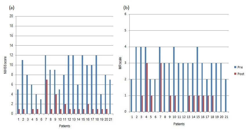
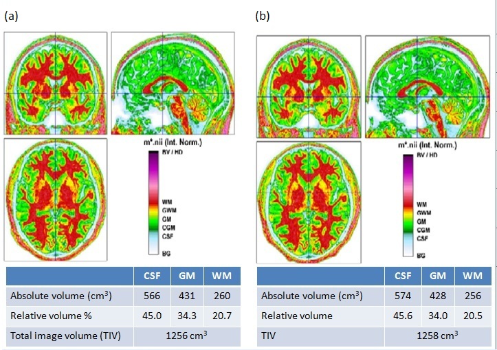
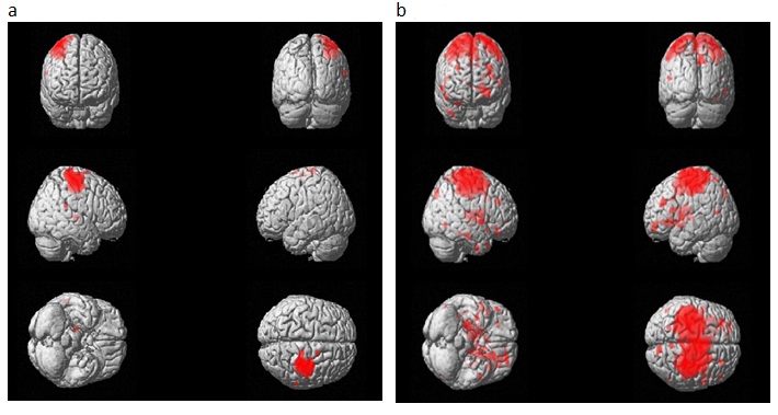
Figure 1 shows the NIHSS (Fig 1a) and mRS (Fig 1b) scores pre- and post physiotherapy intervention. These scores are objective measurements of the impairment caused by stroke and their reduction indicate improvement. The segmentation data analysis did not show much changes (Fig. 2): cerebral spinal fluid (+ 0.6%), gray matter volume (- 0.3%) and white matter volume (- 0.2%). Figure 3 shows a representative fMRI data for the bilateral motor task pre- and post intervention. Post-physiotherapy, activation was observed in motor cortex (cerebral parieto-occipital) in both the left and right hemispheres (Fig 3b), which was absent pre-intervention (Fig 3a). This data is from a patient who had a pre-intervention NIHSS score of 8 and a score of 0, three months post-physiotherapy.Conclusion
Physiotherapy is the conventional method for rehabilitation of stroke patients. The current study has evaluated for the first time, the effects of physiotherapy in post-stroke recovery using MRI. The preliminary results from MR studies reflect the recovery indicated by reduced NIHSS and mRS scores. Functional MRI has indicated the regain in motor activity in the affected hemisphere. Though further in-depth studies and analyses are underway, the preliminary results have shown that MRI can be an important technique in understanding and assessing the physiotherapy induced changes in the recovery of stroke patients.Acknowledgements
No acknowledgement found.References
1. World Health Organization, Global Strategy for the Prevention and Control of Non-communicable Diseases. Report by the Director General. A 53/14 Fifty-third World Health Assembly, May 2000.
2. Carvalho R, Azevedo E, Marques P, Dias N, Cerqueira JJ. Physiotherapy based on problem-solving in upper limb function and neuroplasticity in chronic stroke patients: A case series. J Eval Clin Pract. 2018, 3: 552-560.
3. Donaldson C, Tallis RC, Pomeroy VM. A treatment schedule of conventional physical therapy provided to enhance upper limb sensory motor recovery after stroke: expert criterion validity and inta-rater reliablity. Physiotherapy, 2009; 95(2); 110-119.
Figures