2740
Negative BOLD cerebrovascular reactivity in stroke patients: a sign of misery perfusion of the affected hemisphere1University Hospital Zurich, Zurich, Switzerland
Synopsis
Subjects with hemodynamic failure stage 2 (i.e. misery-perfusion) have heightened risk of acute and chronic brain tissue damage. One of the most important signs of misery-perfusion is a negative cerebrovascular reactivity (CVR). CVR is defined as a blood flow response to a vasoactive stimulus. Recently blood-oxygenation-level-dependent (BOLD) CVR was proposed to detect misery-perfusion. However, BOLD-CVR MRI signal does not reflect CBF changes directly and discrepancies between negative BOLD-CVR and negative CBF changes have been reported. To better assess these discrepancies, we performed a multimodal clinical misery-perfusion assessment with perfusion-weighted-MRI and transcranial-Doppler complimentary to BOLD-CVR in patients with symptomatic steno-occlusive disease.
INTRODUCTION
Subjects with hemodynamic failure stage 2 (i.e. misery perfusion) are known to have a heightened risk of acute and chronic brain tissue damage. One of the most important signs of misery perfusion is a paradoxical – negative – cerebrovascular reactivity (CVR). CVR is defined as a blood flow response to a vasoactive stimulus. Recently blood-oxygenation-level-dependent (BOLD) CVR has been proposed to detect misery perfusion. However, BOLD MRI signal does not reflect CBF changes directly and discrepancies between negative BOLD-CVR and negative CBF changes have been reported. To better assess these discrepancies, we opted to acquire a multimodal clinical misery perfusion assessment with perfusion weighted MRI (PW-MRI) and transcranial Doppler (TCD) complimentary to BOLD-CVR in patients with symptomatic steno-occlusive disease. Both modalities are standard of care for stroke patients after the acute ischemic stroke phase and capable of showing different aspects of misery perfusion. Here, we investigate whether regions with negative BOLD-CVR values present within the symptomatic hemisphere are associated with classical hemodynamic signs of misery perfusion and increased need for collateral blood flow. Only regions of negative BOLD-CVR within the symptomatic hemisphere was investigated due to the qualitative nature of PW-MRI, which requires hemispheric comparison rather than quantitative measurements.METHODS
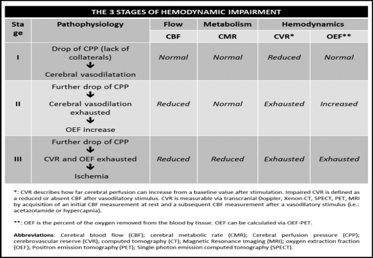
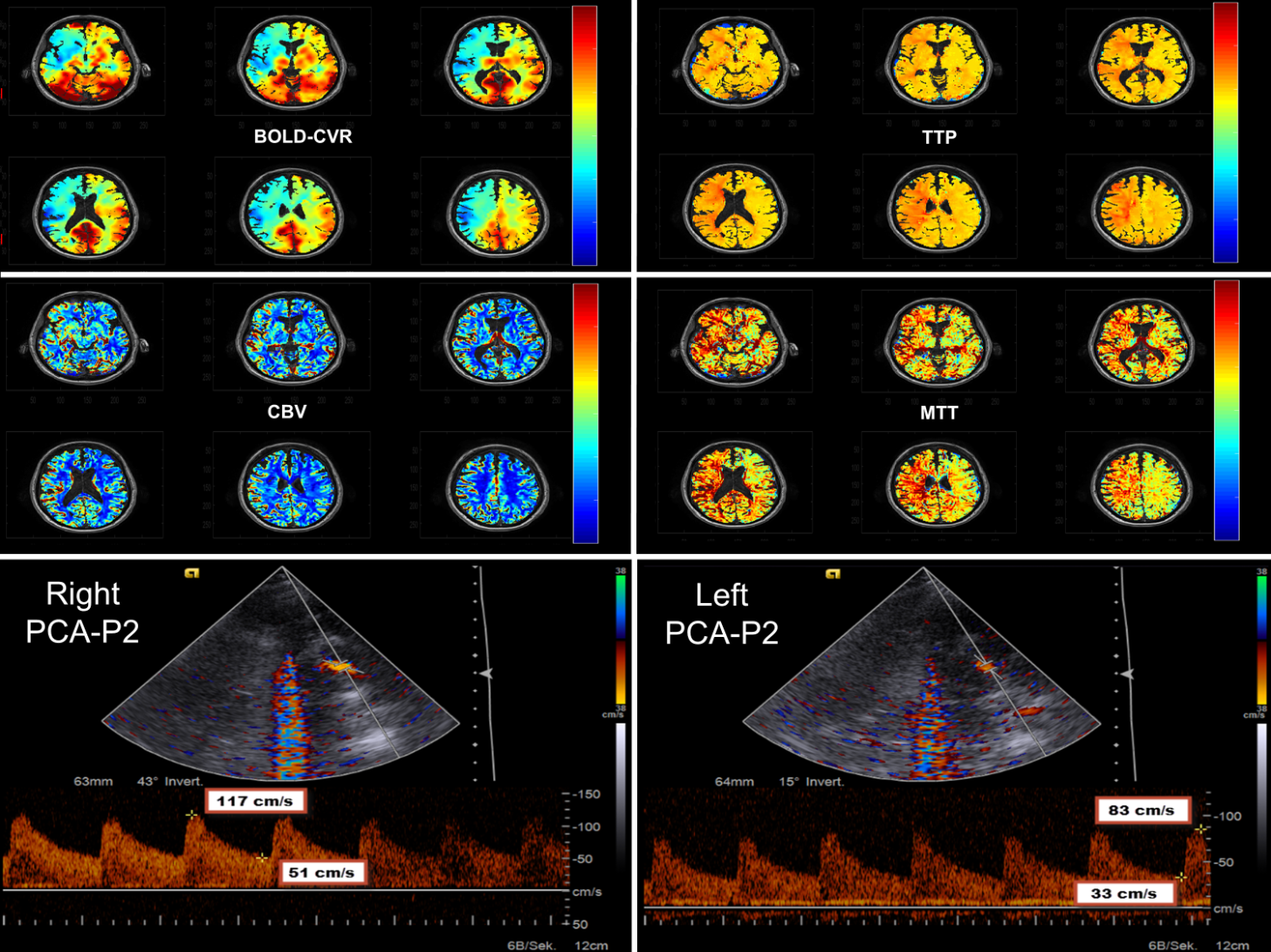
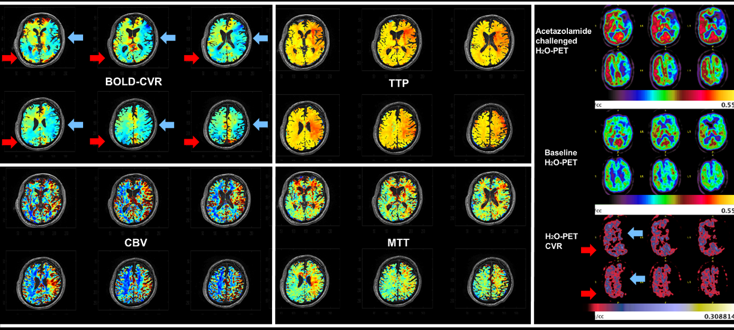
Twenty-one participants with unilateral symptomatic stroke within the anterior circulation and ipsilateral brain regions with negative BOLD-CVR, who also underwent Perfusion-weighted (PW)-MRI and transcranial doppler (TCD) were included. Further details on the setup have been described in previous publications1, 2. Iterative temporal decomposition of the BOLD-CVR data was used to avoid transient phases confounds. The region of negative CVR was extracted and used as a region of interest on the PW-MRI-weighted images (cerebral blood flow, cerebral blood volume, time-to-peak and mean transit time. As comparison, analysis contralateral analysis by flipping the region with negative BOLD-CVR was done. Flow velocities of the posterior circulation (PCA) of the ipsi- and contralateral hemisphere were compared. Last, the volume of negative BOLD-CVR and mean BOLD-CVR within the region with negative BOLD-CVR were correlated to PW-MRI derived parameters and TCD findings.RESULTS
Mean transit time and time-to-peak were significantly prolonged within the region of negative BOLD-CVR in the ipsilateral hemisphere. Cerebral blood volume was also significantly increase, whereas cerebral blood flow did show a difference dependent upon the time delay between the stroke event and the MRI data acquisition – as reported in the literature. The flow velocity within the ipsilateral PCA-P2 segment was also significantly increased and correlated strongly with the volume of negative BOLD-CVR and mean BOLD-CVR within this region.DISCUSSION & CONCLUSION
Here we have shown that brain regions with negative BOLD-CVR present within the ipsilateral symptomatic hemisphere exhibit classical hemodynamic sign of misery perfusion and increasing need for compensatory leptomeningeal collateralisation. Specifically, regions with negative BOLD-CVR show an increase in CBV, as well as a prolonged MTT and TTP.3-5 The only parameter not showing classical signs of misery perfusion is CBF, where one would expect . Here CBF did not show a significant difference between ipsi- and contralateral hemisphere. Regarding collateralisation, the diastolic and systolic PCA-P2 flow velocity was significantly increased towards the ipsilateral hemisphere indication a need for compensatory collateral pathways to preserve perfusion.6 This can also be seen with the strong correlation between volume of the region of negative BOLD-CVR, the average CVR within the region of negative BOLD-CVR and the systolic PCA-P2 flow. With increasing volumes and decreasing quantitative negative CVR, the need for compensatory flow over the PCA-P2 will increasing.
Brain regions with negative BOLD-CVR present within the ipsilateral symptomatic hemisphere exhibit classical hemodynamic sign of misery perfusion and increasing need for compensatory leptomeningeal collateralisation, increasing the evidence of a true vascular phenomenon.
Acknowledgements
No acknowledgement found.References
1. van Niftrik CHB, Piccirelli M, Bozinov O, Pangalu A, Fisher JA, Valavanis A, et al. Iterative analysis of cerebrovascular reactivity dynamic response by temporal decomposition. Brain Behav. 2017;7:e00705
2. van Niftrik CH, Piccirelli M, Bozinov O, Pangalu A, Valavanis A, Regli L, et al. Fine tuning breath-hold-based cerebrovascular reactivity analysis models. Brain Behav. 2016;6:e00426
3. Ferrari M, Wilson DA, Hanley DF, Traystman RJ. Effects of graded hypotension on cerebral blood flow, blood volume, and mean transit time in dogs. Am J Physiol. 1992;262:H1908-1914
4. Schramm P, Schellinger PD, Klotz E, Kallenberg K, Fiebach JB, Kulkens S, et al. Comparison of perfusion computed tomography and computed tomography angiography source images with perfusion-weighted imaging and diffusion-weighted imaging in patients with acute stroke of less than 6 hours' duration. Stroke; a journal of cerebral circulation. 2004;35:1652-1658
5. Grubb RL, Jr., Derdeyn CP, Videen TO, Carpenter DA, Powers WJ. Relative mean transit time predicts subsequent stroke in symptomatic carotid occlusion. Journal of stroke and cerebrovascular diseases : the official journal of National Stroke Association. 2016;25:1421-1424
6. Schneider J, Sick B, Luft AR, Wegener S. Ultrasound and clinical predictors of recurrent ischemia in symptomatic internal carotid artery occlusion. Stroke; a journal of cerebral circulation. 2015;46:3274-3276
7. Esposito G, Amin-Hanjani S, Regli L. Role of and indications for bypass surgery after carotid occlusion surgery study (coss)? Stroke. 2016;47:282-290
Figures