2734
Remote Effect of Ischemic Stroke: Anatomical Specification of Oxygenation Alteration Investigated by Voxel Based R2' Quantification1Shenzhen Institutes of Advanced Technology, Chinese Academy of Sciences, Shenzhen, China, 2University of Chinese Academy of Sciences, Beijing, China, 3Peking University Shenzhen Hospital, Shenzhen, China
Synopsis
Ischemic stroke (IS) may induce oxygenation alterations in brain regions remote to the lesion. Remote effect of IS in terms of oxygen metabolism was evaluated based on the voxel wise R2' quantification for subjects with first ever single lesioned IS in corona radiata (CR) (n=10) and brainstem (n=6) using R2' of the superior sagittal sinus as the reference. Both CR and brainstem IS groups showed significant changes of R2' in distributed brain regions with anatomical specifications, suggesting that IS rather represents a spectrum of pathophysiological events of hemodynamic and metabolic impairments at the global level than a focal vascular failure.
Introduction
Ischemic stroke (IS) may induce extensive hemodynamic and metabolic alterations, which may tightly related to the disease dynamic and recovery [1]. In this study, we aim to investigate the remote effect of IS at the global level in terms of oxygen metabolism based on R2' quantification for subjects with first ever single lesion IS in corona radiata (CR) and brainstem, respectively.Methods
This study was approved by local institutional review board. Ten subjects confirmed with acute IS in CR (m/f=7/3, age ranged 44 to 93 years, averaged 68.5±18.8 years) and six subjects confirmed with brainstem IS (m/f=4/2, age ranged 49 to 77 years, averaged 61.7±10.2 years) were consecutively recruited. T2 and T2* parametric imaging were performed on a 3.0T MR scanner (Siemens MAGNETOM Spectra) using a 12-channel phased-array head coil. Multi-echo turbo spin echo (multi-TSE) sequence was employed for T2 mapping with TR 6500 ms, echo numbers 3, TE (13, 152, 292 ms), slice thickness 3 mm, FOV 178*220 mm, acquisition matrix 256*177, and percent sampling 85%. For quantitative T2* calculation, a fast low angle shot sequence sequence was applied with parameters TR 2500ms, TE (10, 18, 26, 34, 42, 50ms), slice thickness 3 mm, FOV 176*219 mm, acquisition matrix 192*154 and flip angle 30o. The T2 and T2* maps of each subject were spatially aligned with an affine transformation using FSL. R2' was calculated as R2' = 1/T2' = 1/T2*-1/T2. T2 TSE images were spatially normalized to T2 template in MNI space supplied with SPM8 and the normalization parameters were then used to transfer the R2' images. AAL template was used as multi-labeled mask to extract the mean R2' value within each labeled brain area (regions of interest, ROI). The average value of R2' in the superior sagittal sinus (SSS) for each subject was measured as a convenient reference. The R2' difference between each AAL labeled ROI and SSS were explored with Wilcoxon Sign-Rank Test (p<0.05).Results
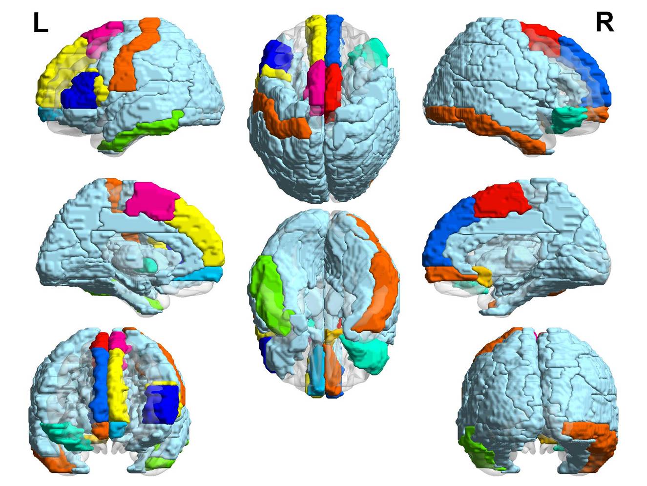
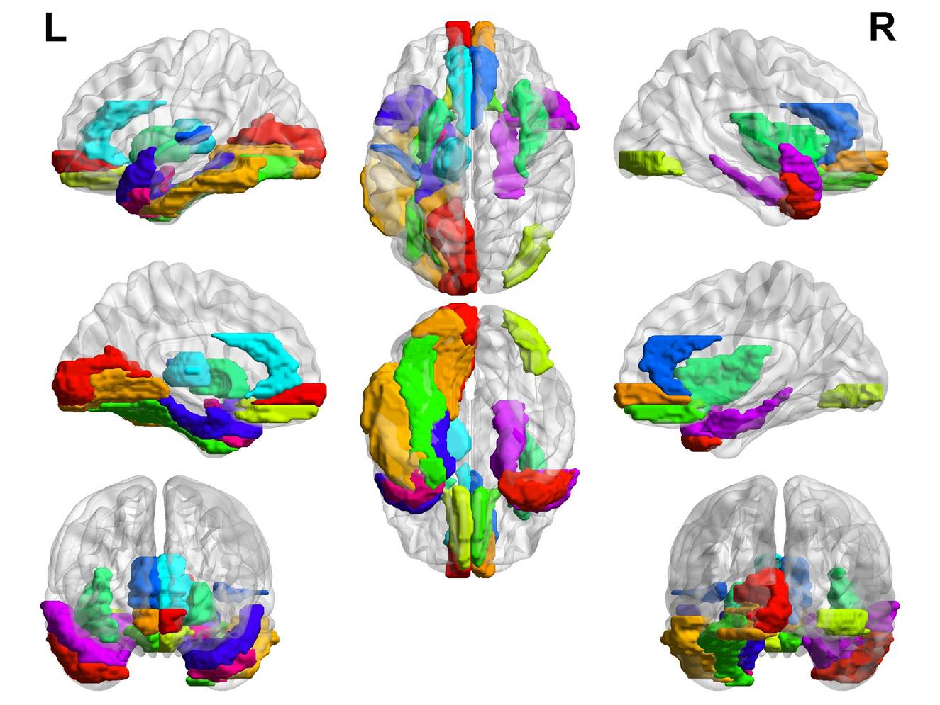
The R2' value of SSS in CR group (43.69±1.89) was significantly higher than that of brainstem IS group (28.26±2.1) (p<0.0001). Referring to SSS, R2' significantly altered in wide spread ROIs for both CR and brainstem IS groups. In CR group, areas mediating somatosensory perception, mood, language and cognitive processing showed significant decrease in R2', mainly including bilateral cingulum and basal ganglia, temporal lobe and bilateral hippocampus. For subjects with brainstem IS, R2' showed significant decrease in bilateral frontal medial orbital gyri, anterior cingulum, right insular, left thalamus and left Heschl gyrus, while increased mainly in left rectus, bilateral parahippocampal, left amygdala, bilateral superior and middle temporal pole and inferior temporal gyrus.Discussion
R2' was proved to be related with oxygen metabolism [2]. SSS has been conveniently used as a reference for the study of cerebral oxygenation in human brain. Both CR and brainstem IS groups showed widespread R2' alteration in this study, suggesting that, rather than vascular failure, focal IS features a global hemodynamic impairment in terms of oxygen metabolism indexed by R2'. This may underlie the various neurological and psychological symptoms in IS subjects. Moreover, the pattern of R2' alteration differs with the location of the lesion, suggesting an anatomical specification in the remote effect of IS.Conclusions
Focal CR or brainstem IS induces extensive impairment in terms of oxygen metabolism indexed by R2' with anatomical specifications. IS may rather represent a spectrum of pathophysiological events of hemodynamic and metabolic impairments at the global level than a focal vascular failure.Acknowledgements
No acknowledgement found.References
[1] Tsivgoulis G, et al. Cerebral hemodynamics in acute stroke: pathophysiology and clinical implications. J of Vascular and interventional Neurology 2008; 1: 65-69.
[2] Seiler A, et al. Oxygenation-sensitive magnetic resonance imaging in acute ischemic stroke using T2'/ R2' mapping: influence of relative cerebral blood volume. Stroke 2017: 48(6):1671-1674.
Figures