2733
Neurodegeneration of the substantia nigra after ipsilateral infarct: quantification with MRI R2* mapping and relationship to clinical outcome1Neuroimaging Dept., Bordeaux University hospital, Bordeaux, France, 2INSERM U1215, University of Bordeaux, Bordeaux, France, 3Neuroimaging Dept., Lille University hospital, Lille, France, 4Division of MRI research, Radiology, Beth Israel Deaconess Medical Center, Harvard Medical School, Boston, MA, United States, 5USMR Dept., Bordeaux University hospital, Bordeaux, France, 6Neurology Dept., Bordeaux University hospital, Bordeaux, France
Synopsis
We tested whether long-term neurodegeneration of substantia nigra (SN) secondary to disconnection by supra-tentorial infarcts can be quantified with iron-sensitive imaging and contributes to clinical outcome. 181 stroke patients (75 striatum infarcts, 106 other locations) were prospectively evaluated at 24-to-72h and at one-year clinically and with MRI to quantify iron through R2*. We showed a delayed increase of R2* within SN that was strongly and independently associated with infarct location along known anatomic projections from SN. Such increase of R2* was an independent contributor of poor motor outcome. Iron-sensitive imaging can monitor neurodegeneration non-invasively within SN and potentially other areas.
Introduction
Long-term clinical outcome after stroke is the result of the infarct itself but may also be impacted by delayed neurodegeneration of remote but anatomically connected areas that become disconnected as a result of the primary ischemic injury1. The substantia nigra (SN) is one region suspected to be affected after infarction, in view of its large array of connections with the supra-tentorial brain2. So far, only transient modifications (ADC decrease) have been measured within SN during the first days following stroke3, 4. To our knowledge, there have been no MRI markers to capture the long-term consequences of stroke on the SN and therefore the clinical impact of such remote alterations is still unknown. In pathological studies, excess of iron has been associated with neurodegeneration; iron being released from dying neurons5. Therefore we hypothesized that R2*mapping could capture the long-term remote degeneration of SN in order to test the association between these changes and stroke outcome.Methods
Patients admitted with a diagnosis of supra tentorial infarct sparing the SN were prospectively evaluated at 24-to-72h (baseline) and at one-year clinically and with MRI to quantify R2*. R2* maps were first inspected for quotation of asymmetry between right and left SN using a Likert-type scale. The SN was then segmented bilaterally to calculate an R2* asymmetry index (SN-AI). We focused on the 95th percentile of R2* (SN-AI95) as a metric of high iron content. SN-AI95 was compared according to infarct location with unpaired t-test and also regressed with variables expected to influence iron accumulation. Voxel based analysis was conducted on average R2* maps6. We also identified individual voxels whose infarction was significantly associated with high SN-AI95 through voxel-based lesion-symptom mapping (VLSM)7. Multivariable regression models were used to test the independent association between SN-AI95 and clinical scores.Results
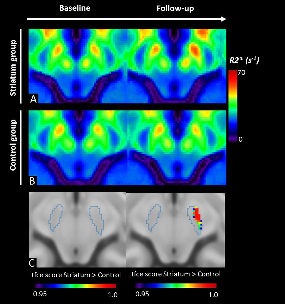
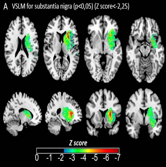
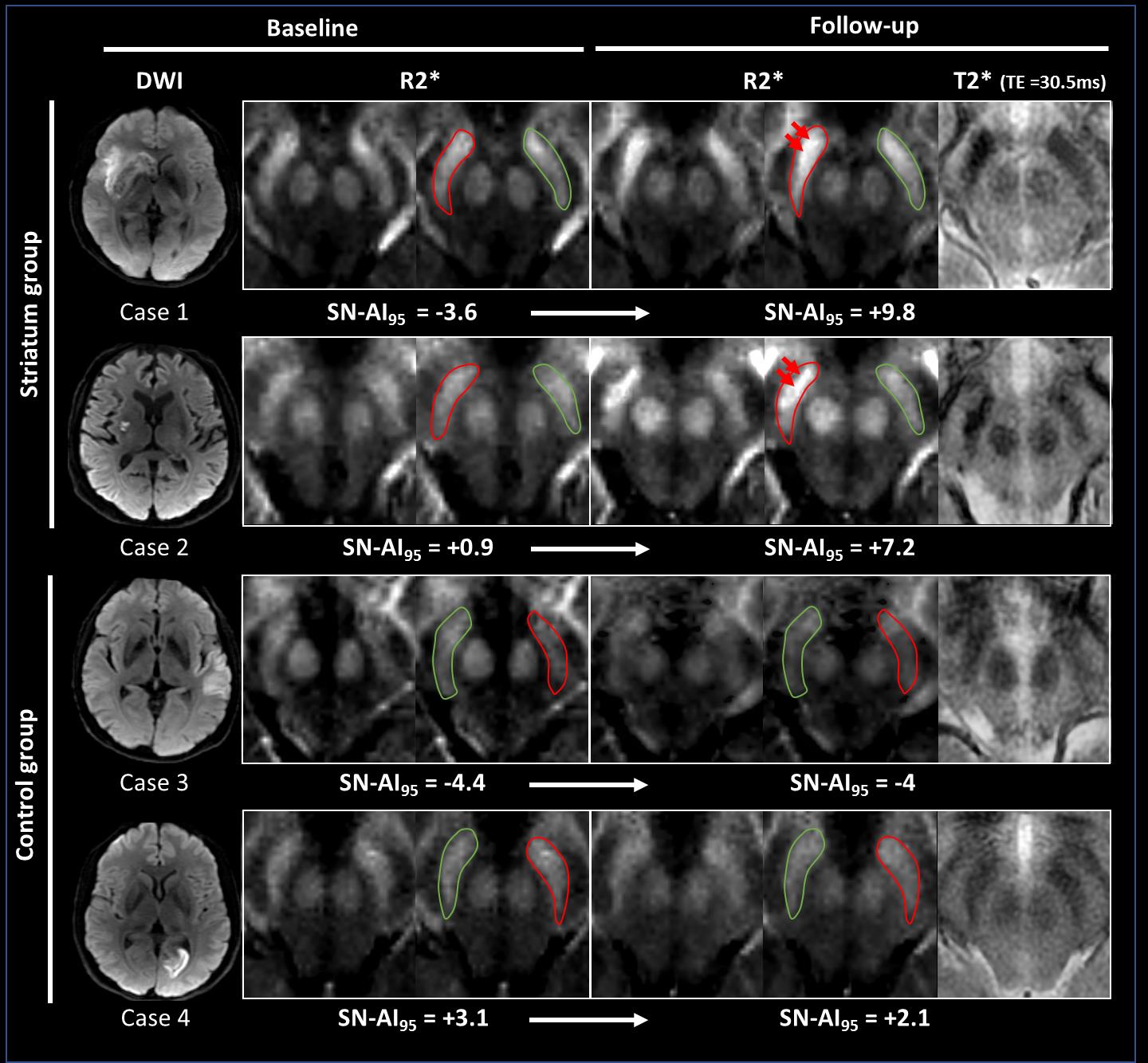
Of 181 stroke patients, the striatum was involved in 75 patients and it was not in 106 patients (controls). Visual inspection of R2* maps identified obvious area of high R2* within the SN ipsilateral to the infarct in 76% of patients with infarct involving the striatum and in 4% of control patients. Illustrative cases also showed that brighter R2* spots could be observed within the lateral part of SN (Figure 1). Quantitative data from masks of SN showed no modification of SN-AI at baseline but significant increase at 1 year that was driven by patients from the striatum group who showed higher SN-AI than control patients (p<0.0001, Figure 2). Average R2* maps within the MNI space confirmed the delayed increase of R2* ipsilateral to infarct when striatum was involved. The voxel-based analysis also confirmed the visual inspection by identifying significant increase of R2* only within the lateral part of SN (Figure 3). This association was independent of infarct volume, baseline SN-AI95 and other confounders (β=4.99 [2.94; 7.04], p< 0.001). We also aimed at mapping the most significant locations at the voxel level instead of considering only the a priori dichotomy based on striatum involvement. The VLSM maps confirmed the strong association between striatum infarction and significant increase of SN-AI95 at follow-up but also identified infarcts involving the insula, the internal and external capsules as significantly associated with increased SN-AI95 at follow-up (Figure 4).
In multivariable regression models, we found that such increase of SN-AI95 was an independent contributor of poor motor outcome but not of cognitive or emotional outcome.
Discussion
We showed a delayed increase of R2* within SN ipsilateral to infarcts which we interpret as iron accumulation associated with long-term remote neurodegeneration. By doing so, we could demonstrate that degeneration of SN significantly worsens clinical outcome independently from the remote infarct. Interestingly, R2* was spatially heterogeneous and mainly increased within the lateral part of SN in relation with the vulnerability of this portion to the GABA/glutamate imbalance8. SN has been recently parcellated based on a tripartite connectivity with limbic (medial), cognitive (ventral) and motor (lateral) arrangements2. In agreement with this parcellation, the predominantly lateral alterations were associated with motor impairment but not with cognitive or emotional performances.Conclusion
R2* can quantify long-term secondary neurodegeneration of SN remotely from an infarct. This neurodegeneration may impact clinical outcome. This finding paves the way toward utilization of iron-sensitive imaging to monitor neurodegeneration non-invasively within SN or other areas9.Acknowledgements
The study was supported by public grants from the French Agence Nationale de la Recherche within the context of the Investments for the Future Program, referenced ANR-10-LABX-57 and named “TRAIL” (Translational Research and Advanced Imaging Laboratory). The study was funded by a public grant from the French government (PHRC protocole hospitalier de recherche clinique inter-régional) funded in 2012.References
1. Zhang J, Zhang Y, Xing S, Liang Z, Zeng J. Secondary neurodegeneration in remote regions after focal cerebral infarction: A new target for stroke management? Stroke. 2012;43:1700-1705
2. Zhang Y, Larcher KM, Misic B, Dagher A. Anatomical and functional organization of the human substantia nigra and its connections. Elife. 2017;6
3. Nakajima M, Inatomi Y, Okigawa T, Yonehara T, Hirano T. Secondary signal change and an apparent diffusion coefficient decrease of the substantia nigra after striatal infarction. Stroke. 2013;44:213-216
4. Winter B, Brunecker P, Fiebach JB, Jungehulsing GJ, Kronenberg G, Endres M. Striatal infarction elicits secondary extrafocal mri changes in ipsilateral substantia nigra. PLoS One. 2015;10:e0136483
5. Thomsen MS, Andersen MV, Christoffersen PR, Jensen MD, Lichota J, Moos T. Neurodegeneration with inflammation is accompanied by accumulation of iron and ferritin in microglia and neurons. Neurobiol Dis. 2015;81:108-118
6. Peran P, Hagberg G, Luccichenti G, Cherubini A, Brainovich V, Celsis P, et al. Voxel-based analysis of r2* maps in the healthy human brain. J Magn Reson Imaging. 2007;26:1413-1420
7. Bates E, Wilson SM, Saygin AP, Dick F, Sereno MI, Knight RT, et al. Voxel-based lesion-symptom mapping. Nat Neurosci. 2003;6:448-450
8. DeGiorgio LA, Dibinis C, Milner TA, Saji M, Volpe BT. Histological and temporal characteristics of nigral transneuronal degeneration after striatal injury. Brain Res. 1998;795:1-9
9. Kuchcinski G, Munsch F, Lopes R, Bigourdan A, Su J, Sagnier S, et al. Thalamic alterations remote to infarct appear as focal iron accumulation and impact clinical outcome. Brain. 2017;140:1932-1946
Figures
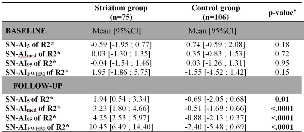
Substantia nigra asymmetry index (SN-AI) of the parameters of R2* histogram within both groups at baseline (24-to-72h) and at follow-up (1 year).
SN-AI5, asymmetry index of the 5th percentile of R2*;
SN-AImed, asymmetry index of the median of R2*;
SN-AI95, asymmetry index of the 95th percentile of R2*;
SN-AIFWHM, asymmetry index of full-width at half-maximum of R2*.