2730
Using Vascular Territories to Predict Disconnection Profiles in Post-Stroke Aphasia1Neuroscience and Aphasia Research Unit, Division of Neuroscience and Experimental Psychology, School of Biological Sciences, University of Manchester, Manchester, United Kingdom, 2MRC Cognition and Brain Sciences Unit, University of Cambridge, Cambridge, United Kingdom, 3Department of Psychology, University of Cambridge, Cambridge, United Kingdom, 4Quantitative Biomedical Imaging Laboratory, Division of Neuroscience and Experimental Psychology, University of Manchester, Manchester, United Kingdom, 5Bioxydyn Ltd., Rutherford House, Manchester, Manchester, United Kingdom
Synopsis
Damage sustained to the brain post-stroke appears random but it may be constrained by the underlying neurovasculature; brain regions supplied by the occluded arterial branch will be affected. Combinations of vascular territories were matched to lesions from 62 post-stroke patients. Anatomical connectivity mapping, a measure of whole-brain connectivity, was used to estimate disconnection in each patient through summing disconnection associated with the territories which best matched their lesion. This novel methodology demonstrated that disconnection following a left-hemispheric stroke can be explained by the underlying neurovasculature and may be of particular interest when no diffusion data is available in the patient.
Introduction
Disconnection patterns in white matter are important for understanding disorders of higher-order functions1. However, as diffusion data suitable for tractography are rarely collected clinically, recent studies have attempted to predict disconnection patterns using other structural aspects such as lesion location. Often these studies overlay lesions onto atlases of white matter tracts to estimate probable disconnection2,3, however these methodologies overlook that although damage sustained to the brain post-stroke appears random, it may be constrained by the underlying neurovasculature; brain regions supplied by the occluded arterial branch will be affected. If a lesion is dependent on the underlying neurovasculature, understanding which vascular territories were damaged may yield a useful way to predict disconnection patterns. Consequently, the aims of this study were; (a)to identify disconnection profiles associated with damage in the middle cerebral artery (MCA) vascular territories (b)to determine whether these vascular territories can be combined to ‘build’ a lesion, and (c)to predict disconnection patterns in patients by the summation of disconnection associated with each damaged vascular territory.Methods
Diffusion-weighted data was acquired using a Philips 3T Achieva in 62 individuals with a left-hemispheric stroke using a pulsed gradient spin-echo planar imaging sequence implemented with TE=54ms, 112x112 image matrix reconstructed to 128x128, reconstructed in-plane resolution 1.875x1.875 mm2, slice thickness 2.1mm, 60 contiguous slices, 43 non-collinear diffusion sensitisation directions at b=1200s/mm2, 1 at b=0. Automated lesion identification algorithms were used to identify the lesion. Combinations of vascular territories were used to find the best match to the lesions (measured using a Jaccard similarity coefficient). Anatomical Connectivity Mapping(ACM) assesses long-range disconnection and may be a complementary alternative to local connectivity measures4. Using probabilistic fibre orientation estimates in FSL, ACMs are obtained by initiating streamlines from all parenchymal voxels. Cumulative trajectories of the streamlines are recorded across all voxels, providing a final brain map indicating how many times streamlines passed through each voxel (i.e. the global connectivity of each voxel). This enables the identification of disconnection outside the damaged tissue as fewer streamlines would be identified at any voxel which was previously connected to the damaged regions. Therefore, predictions of disconnection may be sensitive to widespread damage away from the lesion. It also allows for ‘pseudo-lesioning’ which involves the selective removal of certain brain regions from healthy controls. These ‘pseudo-lesioned’ ACMs can be directly compared back to the whole ACM in the same individual so widespread disconnection following damage to these regions can be ascertained. Through the selective removal of each vascular territory as identified in a previous study5, disconnection profiles were calculated for each territory, reflecting where connectivity was lower when a territory was removed. ACM was used to estimate the disconnection in each patient through combining the disconnection patterns associated with each territory which best matched their lesion. This was compared back to the actual ACM disconnection patterns found within the patient.Results
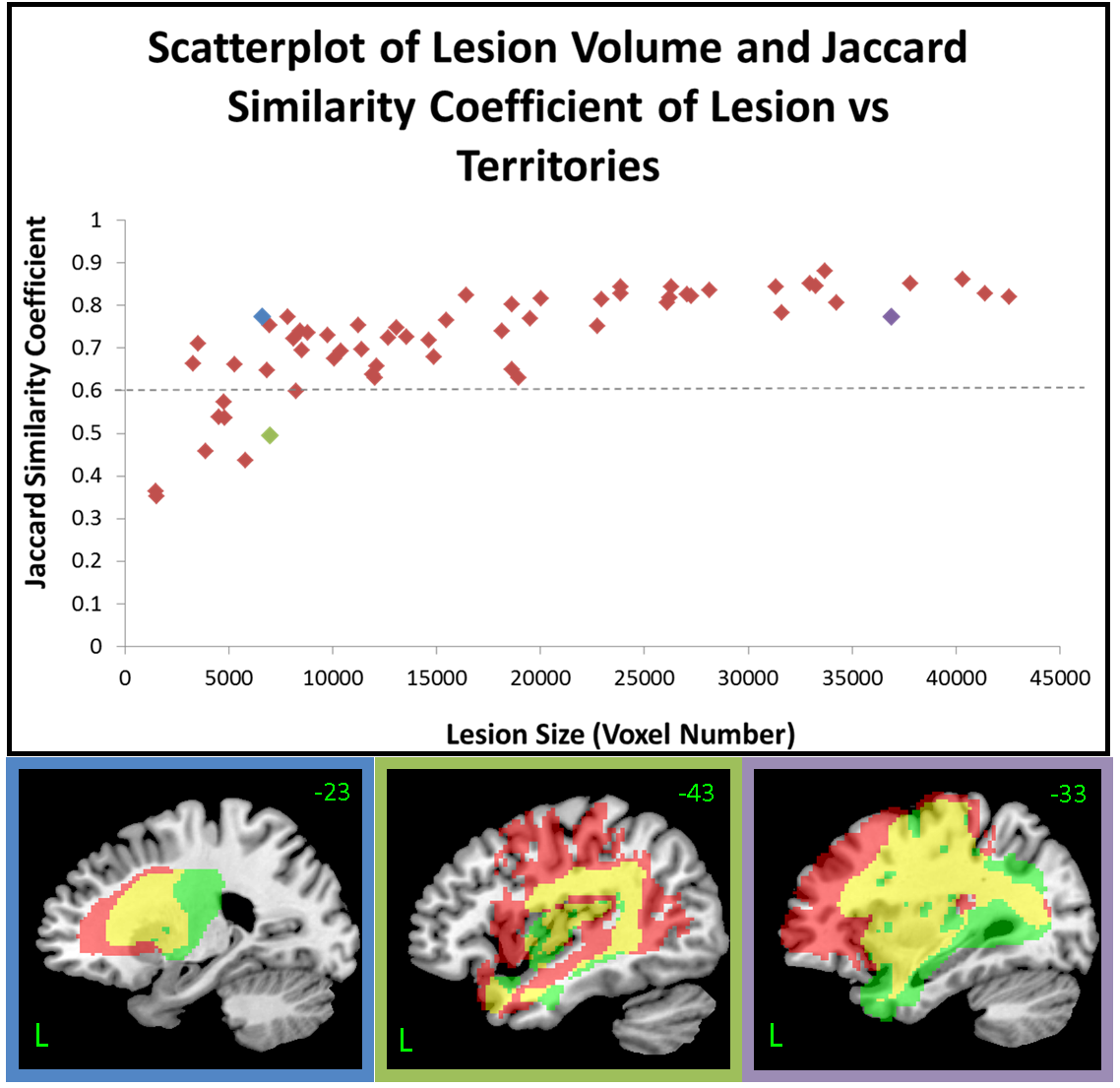
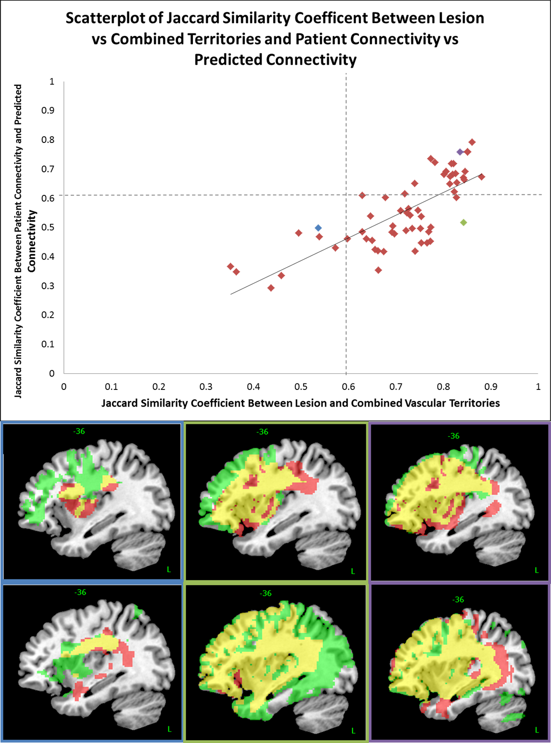
The selective removal of each vascular territory through pseudo-lesioning revealed disconnection associated with damage to each territory. The mean number of territories combined to match the lesion was 4.58. The average Jaccard similarity score between the lesion and the best combination of territories was 0.72(0.6 is considered high). A significant positive Pearson’s correlation was found between lesion volume and Jaccard score(r=0.665, p<0.001). The average Jaccard score between actual and predicted disconnection in patients was 0.56. A higher similarity between the lesion and territories was significantly positively associated with a closer match between the predicted and actual patient connectivity. Lesions smaller than 10,000 voxels (mean=0.45) had a significantly lower similarity between predicted and real connectivity then larger lesions (mean=0.61), t(62)=7.72, p<0.001).Discussion
A striking result was that profiles revealed disconnection which extended far beyond the removed region. The close match between combined vascular territories and the lesion for each individual confirms that the underlying neurovasculature can explain the remote damage sustained following a left-hemispheric MCA stroke. In addition, high similarity scores were found between predicted and real connectivity scores in the patients suggests that connectivity can be predicted using the underlying neurovasculature alone and may be more accurate than predictions using only local diffusion measures.Conclusion
This novel methodology of pseudo-lesioning demonstrated that disconnection following a left hemispheric stroke can be explained by the predicted underlying neurovasculature of the MCA alone, to a high degree of accuracy. By using these vascular territories, disconnection associated with a lesion can be predicted. This may be of particular interest when there is no scope for the collection of tractography-suitable diffusion data in the patients themselves and may aid in understanding the behavioural deficits that patients may present post-stroke.Acknowledgements
Data were partially collected by Dr Rebecca Butler and Blanca De Dios. PCA factor score loadings were provided by Ruth Ingram. We are grateful to all participants and carers for their contribution in this research study. NB was supported by an iCASE MRC studentship in collaboration with Bioxydyn Ltd. ADH was supported by funding from the Rosetrees Trust. MALR was supported by an MRC programme grant (MR/J004146/1) and an ERC advanced grant (GAP: 670428 - BRAIN2MIND_NEUROCOMP).References
1. Wernicke, K. (1874). The aphasia symptom-complex. (J. Benjamins, Trans.) Eling P, editor. Reader in the history of aphasia. (Vol. 4, pp. 69–89).
2. Kümmerer, D., Hartwigsen, G., Kellmeyer, P., Glauche, V., Mader, I., Klöppel, S., . . . Saur, D. (2013). Damage to ventral and dorsal language pathways in acute aphasia. Brain, 136(2), 619-629.
3. Thiebaut de Schotten, M., Dell'Acqua, F., Ratiu, P., Leslie, A., Howells, H., Cabanis, E., . . . Catani, M. (2015). From Phineas Gage and Monsieur Leborgne to H.M.: Revisiting Disconnection Syndromes. Cerebral Cortex (New York, NY), 25(12), 4812-4827. doi:10.1093/cercor/bhv173
4. Embleton, Morris, D. M., Haroon, H. A., Lambon Ralph, M. A., & Parker, G. J. (2007). Anatomical connectivity mapping. Proc Intl Soc Mag Reson Med15.
5. Zhao, Y., Halai, A.D., Lambon Ralph, M.A. (2018) Mapping Both Lesion and Behaviour Structures in Post Stroke Aphasia [in revision]
Figures